
Heat vs Cold Therapy: Which One to Use and When
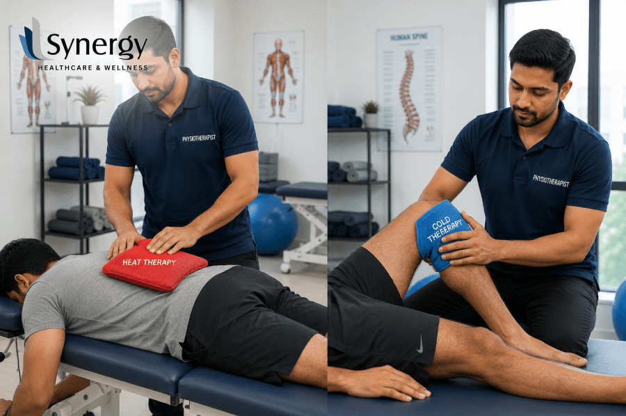
You can take thermotherapy or targeted cold therapy; either way, your recovery path can be drastically different. When to use soothing thermal therapy for muscle stiffness and when to use intense cold compression therapy for acute swelling. This comprehensive guide follows professional guidelines to help you heal effectively, reduce pain and restore mobile health. Introduction: The Science Behind Temperature Therapy for Pain Relief Our natural instinct is to seek immediate relief when physical injury strikes or chronic pain flares up. Two of the most accessible, cost-effective and powerful tools at our disposal are temperature-based modalities: heat and cold. With so many options available, making the right choice can be difficult. Applying an ice pack may lock a stiff, aching muscle into a deeper spasm, while a hot water bottle may cause a fresh, throbbing sprain to pour out inflammatory fluids, thereby increasing local swelling. The trick to knowing when and how to use these tools is to understand how our physiological systems react to changes in temperature. Knowing the exact therapeutic windows for slowing your metabolism with cold therapy or relaxing your muscles with heat treatment can empower you to take an active role in your injury management. We’ll look at the science behind temperature-based recovery to help you choose the best treatment method for your physical condition. How Cold Therapy Works – Calming the Storm To appreciate the importance of the choice of temperature, we must understand the cellular change occurring with the application of cold to living tissue. Cold application works by lowering local metabolic demands, slowing nerve conduction, and reducing blood vessel diameter, thereby limiting tissue activity. Fluid Management and Vasoconstriction When you apply ice or a cold wrap to an injured area, the smooth muscles of the blood vessels constrict in response to the sudden drop in temperature. This process is called vasoconstriction, and it severely limits blood flow locally. When physical trauma occurs, such as a twisted ankle or a torn ligament, the nearby capillaries rupture and leak fluid into the surrounding space, causing swelling and bruising. Ice therapy can immediately halt this over-leaking of fluid, limiting the extent of internal swelling. Diminishing the Pain Signal Besides fluid control, extreme cold acts as a mild local anaesthetic. It reduces the speed at which peripheral nerves transmit pain signals to the central nervous system. The powerful drug-free numbing effect blocks these rapid pain pathways and helps break the intense pain-spasm cycle that is common in fresh injuries. The Benefits of Cold Compression Therapy For extensive swelling or major joint trauma, conventional ice packs often provide limited benefit since they cannot mould evenly around the joint. That’s where Cold and compression therapy come in. This technique eliminates the stagnant fluid in the injured zone and at the same time cools the deep tissue layers by means of active pneumatic compression combined with continuous cooling, thus better controlling the inflammation. Science of Thermotherapy: Restoring Flow and Flexibility Cold is about restriction and calming; thermal therapy is about acceleration, expansion and tissue restoration. When you introduce heat, the vascular network knows to open up and bring fresh oxygenated blood to starving, tight tissue. Vasodilation and Nutrition of the Tissues Heat causes the blood vessels to widen, or vasodilation. This influx of blood flow brings with it a rich supply of fresh oxygen, vital amino acids and essential nutrients directly to the sore muscles, accelerating the natural cellular repair process. And, the increased circulation also flushes out accumulated metabolic waste products, such as lactic acid, which can lead to post-exercise soreness and structural fatigue. Loosening Tight Connective Tissues Collagen is a structural protein that is found in abundance in muscles, tendons and ligaments. When tissue is injured or held in one position too long, these collagen fibres become stiff, hard and resistant to movement. Regular application of heat treatment changes the elastic properties of collagen, making the tissues much more pliant and cooperative. Heat is therefore an excellent modality for chronic morning stiffness, deep muscle spasms and warming up tight joints for stretching and exercise. Strategic Timing: When to Freeze, When to Thaw Condition/Injury Category Proposed Treatment Main Physiological Function Best Treatment Duration Acute Sprains & Strains (First 48 hrs) Cold treatment Reduce swelling, stop bleeding, numb sharp pain 15–20 minutes every 2–3 hours Muscle Stiffness (Neck & Back) – Chronic Heat Treatment Increase blood flow, relieve deep muscle spasms, and increase flexibility 20–30 minutes depending on your needs Severe Swelling Post-Exercise Cold and compression therapy Constrict the vessel and push out the fluid buildup 15 minutes after activity Arthritic Joint Pain – Non-inflammatory Therapeutic Heat Enhance joint elasticity. Improve structural stiffness 20 minutes prior to movement Optimising Synergy Rehabilitation Pathways At Synergy, we combine sophisticated clinical modalities with specific, active physical therapy in our approach to injury management. We know that temperature control isn’t a cure but a great catalyst to get your body ready to move and recover functionally over the long term. Our specialist clinicians apply heat therapy and dynamic cold treatment systems to customise recovery for each stage of your physical rehabilitation: 1. Acute Injury Management Our team utilises targeted compression cold therapy during the sensitive 48-hour period post an acute sports injury or joint sprain. This systematic approach treats early-stage inflammation without interfering with the body’s natural tissue repair processes. Learn more about our Sports Injury Rehabilitation services. 2. Management of Chronic Pain & Spasm If you suffer from chronic back pain, stubborn postural tension or severe neck tightness, we use deep-penetrating thermal treatment. Raising tissue temperature helps to reduce muscle guarding and to relax the constant aches that interfere with life. 3. Mobilisation Before Exercise Our movement specialists often apply localised heat to assist with improving joint range of motion prior to guiding you through customised clinical exercises. This allows for therapeutic movements to be much more comfortable and effective. 4. Recovery After Treatment We might use local cooling techniques after deep tissue mobilisations or intensive manual therapy to make the treated area
Diastasis Recti: What It Is and How to Recover Post-Pregnancy
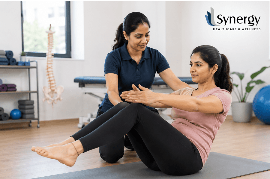
Uncovering what diastasis recti is helps new mothers reclaim their core vitality after childbirth. This article breaks down the diastasis recti meaning, explores the signs of severe diastasis recti, and addresses changes in the post-pregnancy belly. Find out how expert evaluation and customised recovery plans strengthen the abdominal wall and restore functional support without invasive procedures. Introduction: Why Does Your Core Feel Different After Pregnancy? Welcoming a newborn brings immense joy, but it also introduces profound structural changes to a mother’s body. Among the most common physical shifts noticed after childbirth is a persistent softness or bulging in the midline, often referred to as a post-pregnancy belly. While many expect the abdomen to bounce back naturally, a large percentage of women experience a distinct physical condition where the parallel bands of the stomach muscles remain separated. Understanding what is diastasis recti the first crucial step toward effective postpartum healing. It’s not just cosmetic; it’s a structural change in the anterior trunk that can fundamentally change how your body transfers weight and manages internal pressure. Discover the real diastasis recti meaning, how to recognise severe diastasis recti, and how to find precise clinical pathways to safely rebuild your core, preserve your pelvic health, and eliminate discomfort. The True Anatomy: What Does Diastasis Recti Mean? To treat this condition, one needs to study the fundamental muscular architecture of the human torso. The rectus abdominis, better known as the “six-pack” muscle, is made up of two parallel vertical sheets of muscle connected down the middle by a thick band of collagen-rich connective tissue called the linea alba. The uterus steadily increases in size during pregnancy and exerts a constant, progressive outward pressure on the abdominal wall. Hormonal changes (notably the release of relaxin and progesterone) relax the connective tissue throughout the pelvis and torso to allow for this expansion. This pulls the linea alba apart laterally, increasing the distance between the left and right halves of the rectus abdominis muscle. The abnormal wide and thin linea alba is the exact clinical diastasis recti definition, resulting in an inter-rectus distance (IRD) usually more than 2 to 2.5 centimetres in the postpartum period. What is important to realise is that the muscle tissue itself has not torn or ruptured, but the central connective tissue bridge has lost its resting tension and mechanical stiffness, which alters the stability of the whole core system. Recognising the Signs: Mild Separation to Severe Diastasis Recti Many cases of mild separation resolve spontaneously in the first eight to twelve weeks postpartum as hormonal levels normalise and tissues regain elasticity. But if the linea alba is damaged for longer than this time, then there are some visual and functional symptoms to look out for. Mild to Moderate Cases: In most cases, the mother will notice a soft, jelly-like substance around her belly button. When you try to sit up from a lying position, you may see a temporary appearance of a slight ridge or small mound in the middle of the abdomen. This is often associated with a sensation of the lower back working harder than normal during everyday movements. The Effects of Severe Diastasis Recti Diastasis recti is considered severe when the inter-rectus distance is significantly greater than 3 to 4 centimetres or when the underlying fascia is exceptionally thin and compliant. In these advanced cases, the fascial wall is no longer able to support the intra-abdominal organs adequately. When searching for clinical guidance or studying pictures of advanced diastasis recti, patients will observe a highly pronounced, structural protrusion. Rather than a minor ridge, the midline visibly domes, “cones”, or projects outward into a sharp, tent-like shape during tasks that increase intra-abdominal pressure—such as lifting a baby, coughing, or performing an abdominal crunch. In a resting, standing posture, this significant lack of muscular support causes the internal organs to press outward, creating a persistent dome shape that makes the individual look several months pregnant long after giving birth. Why Does a Post-Pregnancy Belly Alter Structural Movement? The visual changes of a postpartum belly are intrinsically linked to deeper mechanical adaptations within the body. The anterior abdominal wall does not function in isolation; it works as part of an integrated canister composed of the diaphragm at the top, the pelvic floor muscles at the base, the multifidus along the spine, and the transversus abdominis wrapping around the waist like a corset. When a significant gap persists along the linea alba, this entire pressure system becomes compromised. Without the proper tension of the centre, the core muscles are not able to generate enough force to stabilise the pelvis and low back. It causes a number of other problems, like: Reclaiming Your Core with Synergy Clinical Expertise Recovering from abdominal separation requires a highly methodical, individualised approach that prioritises neuromuscular control over absolute load. Random core exercises found online can often exacerbate the separation by forcing the superficial obliques to pull the abdominal halves further apart. Safe Postpartum Rehabilitation at Synergy At Synergy, our dedicated women’s health physiotherapists offer customised, evidence-based treatment plans to help restore abdominal strength and function according to the extent of your abdominal separation. In addition to the attempt at closing the gap horizontally, the new goal in modern medicine is to bring functional tension back to the linea alba, thus making sure that your core can effectively transfer its weight without sagging or doming. The path to recovery in Synergy follows a gradual process: 1. Re-training Diaphragmatic Breathing The first stage entails retraining the breathing pattern in order to control intra-abdominal pressure. Deep, lateral costal breathing helps activate the deep core without forcing stressful outward pressure against a healing linea alba. 2. Isolated Transversus Abdominis Activation Using targeted abdominal drawing-in manoeuvres, clients learn to gently contract the deepest layer of the abdominal wall. This exercise acts as a splint by bringing the two sides of the rectus close to each other and tensioning the fascia of the midline. 3. Co-contraction of the Pelvic Floor Pelvic floor co-contraction
Posture and Hygiene in Pregnancy
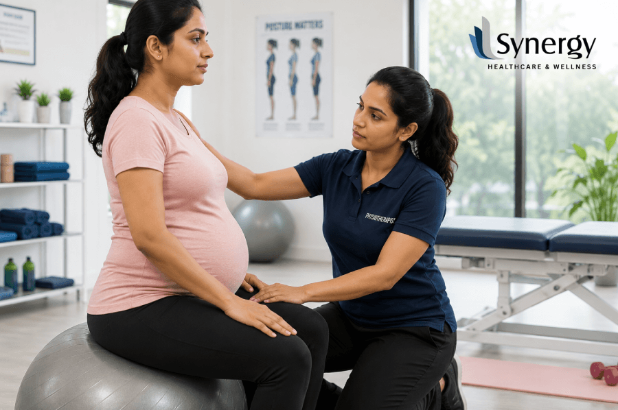
Looking for ways to sit on the floor comfortably during pregnancy or correct the sitting position in pregnancy? Discover safe sitting and sleeping positions, posture tips, and pregnancy do’s and don’ts recommended by physiotherapists to help you stay comfortable. Introduction: Support Your Body Through Every Stage of Pregnancy Pregnancy changes just about everything about how your body moves, balances, and rests, and posture is often the first thing to go. Everyday habits such as crossing your legs or slumping on the sofa can subtly add strain to your back, hips and pelvis as your centre of gravity shifts forward with the growing uterus. One of the simplest ways to relieve discomfort and support a smoother pregnancy is to get the sitting position in pregnancy right as well as good sleeping posture during pregnancy. This guide covers sitting posture during pregnancy, safe ways to sit on the floor during pregnancy, positions to avoid during pregnancy and how the position of the uterus during pregnancy affects the advice doctors give. We’ll also cover general dos and don’ts during pregnancy and how expert physio support, like that provided at Synergy, can help you move through pregnancy with greater comfort and confidence. The Importance of Good Posture During Pregnancy With each passing month of pregnancy, hormones such as relaxin soften the ligaments that hold your joints in place, and your enlarging belly forces your lower back into a deeper arch. These factors increase the vulnerability of the spine and pelvis to the strain of everyday positions that used to seem harmless. Good posture during pregnancy isn’t about looking a certain way. It’s about training your body to stand, sit and lie in positions that cause the least possible stress to your back, hips and pelvic joints. When poor posture is maintained over weeks, it can lead to: Getting the Sitting Position in Pregnancy Right Correct sitting posture while pregnant is about supporting your spine and keeping your pelvis level, not tucked under or slumped. Physiotherapy recommendations typically recommend the following changes: Positions to Steer Clear of Sitting During Pregnancy As part of a wider list of positions to avoid in pregnancy, there are some sitting habits that are best avoided, particularly from the second trimester onwards: Location Why should it be limited Sitting cross-legged for long periods May decrease circulation and increase imbalance in the pelvic joint Slouching or slumping over the bump Causes more upper back strain and less room for a baby Sitting on very low seats or unsupported seats Encourages a tucked pelvis and a rounded spine Sitting for long, unbroken periods Causes stiffness and swelling in the legs Later pregnancy (lying flat on your back for long periods) May promote less favourable foetal position None of these positions are dangerous from time to time – the key is to avoid long, repeated exposure to any one posture. Safe Ways to Sit on the Floor During Pregnancy Sitting on the floor is an everyday part of life, religious activities, or relaxation, and many pregnant women ask how to sit during pregnancy on floor without straining. Floor sitting can still be comfortable well into later pregnancy with a couple of tweaks: Options for Not Sitting Cross-Legged on the Floor If your crossed legs are not comfortable, try the following: If you have symphysis pubis or pelvic girdle pain, it’s usually best to avoid sitting cross-legged on the floor for long periods, but a physiotherapist can suggest suitable alternatives. Pregnancy Sleeping Position: What the Research Shows Sleeping position in pregnancy is one of the most researched topics in prenatal care, and the rationale is more than comfort advice; it is physiological. By about the 20th week of pregnancy, lying flat on your back can cause the growing uterus to compress the large vein that carries blood back to the heart, the inferior vena cava, and sometimes the aorta as well. This can result in decreased cardiac output and uterine blood flow, termed aortocaval or supine hypotensive syndrome. Several case-control studies from New Zealand, Australia and the UK have shown that sleeping on the back in later pregnancy is associated with increased risk of late stillbirth compared with side-sleeping, thought to be related to this reduced uterine perfusion. This evidence has led to the recommendation by national guideline bodies that pregnant women sleep on their side after 28 weeks of gestation. Practical Sleep Positioning Tips Position of the Uterus in Pregnancy and Its Importance for Comfort Understanding the position of the uterus during pregnancy helps explain why advice about posture changes as pregnancy progresses. During uterine enlargement, the uterus transforms from a small pelvic organ to a large abdominal organ that can, in the recumbent position, exert pressure on large blood vessels and, in the chair-slumped position, restrict the movement of the ribs and diaphragm. Keeping a well-supported, upright sitting position and side-lying sleeping posture can help to keep pressure off of these structures and may also help to maintain optimal positioning of the baby as term approaches, as a persistently slumped or reclined posture can reduce the space available for the baby to settle head-down. Want to improve your posture and core stability? Read our blog, Daily Core Strengthening Routine for Better Posture and Balance, for simple exercises and expert physiotherapy tips. Quick Guide: Do’s and Don’ts of Pregnancy Do Don’t Do Sit with your back against a support, with your feet flat Sit or slump for prolonged periods without a break 28 weeks sleeping on your side (preferably the left) Sleep on your back for extended periods of time Sit on the floor with the support of cushions Sit cross-legged for long, unbroken periods Change places every 30-45 minutes Stand or sit in one position for hours Bend your knees and hips to lift Hinge forward at the hips with your knees straight Wear a supportive shoe and maintain good standing posture Wear high heels or shoes that are not supportive for long periods Synergy: Personalised Physiotherapy Care for Pregnant Women
Muscle Recovery: DOMS, Stretching & Strength Testing

Post-workout stiffness: What to do? Delayed onset muscle soreness (DOMS) is a normal response to exercise. Active stretching, proper recovery, and muscle strength testing can reduce discomfort, improve performance, and support a safe return to training. At Synergy, physiotherapist-led care helps you recover effectively. Introduction: From Muscle Soreness to Stronger Performance If you’ve ever tried a new workout, added extra weight to the barbell or gone on a longer run than usual, you’ll be familiar with that feeling: a dull, aching stiffness that comes on a day or two later. This is delayed-onset muscle soreness, and understanding the right delayed-onset muscle soreness treatment can be the difference between a smooth recovery and weeks of nagging discomfort. Clinicians also use muscle testing and a muscular strength test to assess the progress of muscle healing and function, in addition to active stretching and other recovery strategies. These approaches, combined with structured muscle therapy, are the basis of safe, evidence-informed recovery. In this guide, we will break down the science that underlies DOMS, the real-world benefits of stretching, how strength assessments are conducted, and how a physiotherapy-led approach, such as the one offered at Synergy Healthcare & Wellness, promotes long-term muscle health. What Is Delayed Onset Muscle Soreness (DOMS)? Delayed onset muscle soreness is a transient phenomenon that usually manifests 24 to 72 hours following unfamiliar or intense physical activity, particularly exercise involving eccentric muscle contractions – movements in which a muscle lengthens while under tension, such as when lowering a weight or running downhill. DOMS was blamed on lactic acid build-up for years. Current research has gone much further than that theory. Microscopic disruption of muscle fibres and the surrounding connective tissue is now seen, followed by a localised inflammatory response which sensitises pain receptors in the fascia. This inflammatory cascade involves signalling molecules such as interleukins and tumour necrosis factor, as well as a transient increase in blood markers such as creatine kinase, which reflects disturbance of muscle fibres. Common DOMS Symptoms The discomfort is usually most noticeable around the 48-hour mark, and it will usually subside within a week or so. DOMS is usually harmless and self-limiting, but the accompanying strength loss and stiffness can increase the risk of injury through compensation if you get back into training too soon, which is why having a well-thought-out delayed-onset muscle soreness treatment plan is so valuable. Delayed Onset Muscle Soreness Treatment: What Really Works Various physiotherapy interventions have been investigated for the management of DOMS with mixed but increasingly clear results. A systematic review of physical therapy methods found measurable benefits in reducing soreness intensity from massage, cryotherapy, active recovery and some contrast methods, but these were inconsistent. Treatment Strategy How It Helps Strength of Evidence Active Recovery (Light Activity) Improves circulation, eases stiffness Massage/hands-on therapy Decreases perceived muscle soreness and tension Cryotherapy (applying cold) Limits secondary swelling (inflammatory) Dynamic stretching May relieve tightness in the preserved range of motion Re-Loading Strength Gradually Restores muscle function and confidence Nutritional support (polyphenols, protein) Facilitates tissue repair In practice, a DOMS treatment plan typically uses a combination of these strategies rather than one in isolation because no single intervention will completely eradicate DOMS. Importantly, while over-the-counter anti-inflammatories may help with short-term relief, they are not generally recommended as a first-line approach, as research suggests they might impede the muscle’s natural adaptive repair process if used repeatedly. A physiotherapist can help you find out which combination fits your training load, muscle group and recovery time. How Functional Stretching May Aid Muscle Recovery? A cool-down or recovery routine often includes active mobility exercises, which use your own muscle contraction to move a joint through its range, rather than relying on an external force. Functional stretching (as opposed to passive or static stretching), which involves the opposing muscle group, is believed to improve flexibility while improving circulation and neuromuscular control. The research on post-exercise stretching is really mixed. Several meta-analyses have found no significant effect of stretching over simple rest on DOMS reduction or speed of strength recovery; some other reviews report that stretching may be beneficial for tendon compliance, local blood flow and a slight reduction in stiffness, even if the soreness scores are not dramatically different. Recent literature generally agrees that stretching is low-risk, accessible and reasonable as part of a wider recovery programme rather than a stand-alone cure for DOMS. Practical Tips on Active Stretching Muscle and Muscular Strength Test: Why It Matters While soreness tells you how a muscle feels, strength testing tells a clinician how a muscle actually performs. A muscular strength test allows physiotherapists to objectively track whether strength has returned to baseline before a patient resumes full training or heavy lifting, a step often skipped in self-managed recovery. The most widely used clinical method is Manual Muscle Testing (MMT), based on the Medical Research Council (MRC) or Oxford Scale, which grades a muscle’s ability to contract against resistance on a 0–5 scale. MRC / Oxford Scale Grade Description 0 No visible or palpable muscle contraction 1 Flicker of contraction, no movement produced 2 Movement possible with gravity eliminated 3 Movement against gravity, no added resistance 4 Movement against gravity and moderate resistance 5 Normal strength against full resistance For more precise tracking, clinicians may use handheld dynamometers, which quantify force output in kilograms or Newtons rather than relying on subjective grading alone. This is particularly useful when monitoring recovery after an injury, post-surgical rehabilitation, or a demanding sports season, since dynamometry can pick up subtle strength deficits that a manual muscle strength assessment might miss. Regular muscle testing throughout a recovery programme helps a physiotherapist decide: How Does Muscle Therapy Speed Up Recovery? Structured muscle rehabilitation goes beyond simple rest. It typically combines hands-on techniques, such as soft tissue release and myofascial work, with guided active mobility exercises, progressive loading, and periodic muscular strength test check-ins. It’s a multi-layered approach to both the discomfort from DOMS and the tissue and neuromuscular changes that cause it. A trained physiotherapist through
Tailbone (Coccyx) Pain: Causes Without Injury & How Physiotherapy Can Help
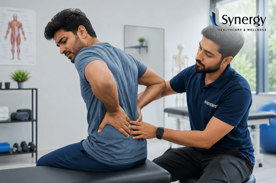
Wondering why your tailbone hurts without an injury? Tailbone pain can result from prolonged sitting, poor posture, arthritis, muscle strain, or cysts. Expert physiotherapy at Synergy Healthcare and Wellness helps relieve pain, improve mobility, and restore everyday comfort. Introduction: Finding the Real Causes of Tailbone Pain Coccyx pain, or tailbone pain, is much more common than most people think, and it isn’t always caused by a fall or an accident. Many people look for tailbone pain causes simply because their discomfort appeared out of nowhere, and they can’t recall any knock or trauma. If you’ve ever asked yourself what causes tailbone pain without injury, you’re not alone – everyday habits, underlying joint changes, and even some medical conditions can quietly trigger pain at the base of the spine. Knowing how the coccyx works, recognising both common and hidden causes of pain, identifying symptoms that need medical attention, and understanding how personalised physiotherapy can support recovery are essential steps towards lasting relief. What Is the Coccyx? A Quick Anatomy Refresher Your coccyx is located at the very bottom of your spine, below your sacrum, and typically consists of three to five small, fused bones. The coccyx is made up of three to five fused vertebrae that support the spine. The coccyx is attached to the sacrum by strong ligaments and is directly attached to several pelvic floor muscles. This is why problems with the coccyx can sometimes affect comfort with the bladder, bowels, or even sex. The tailbone is particularly prone to irritation, even without a single dramatic injury, due to its position, bearing pressure every time you sit, lean back or shift your weight. What Causes Tailbone Pain Without Injury? The classic triggers are a backward fall or childbirth, but a large share of coccydynia cases develops gradually. Understanding what causes tailbone pain without injury can help you get on the right path to treatment sooner. 1. Sitting for a Long Time or Bad Posture Sitting for long periods, particularly in a hard, narrow or non-supporting chair, places continuous pressure on the coccyx. Slouching or leaning back puts additional pressure on this tiny bone, thereby gradually causing inflammation of the ligaments and joints in this area. 2. Ageing and Degenerative Joint Changes The small joints that link the coccyx to the sacrum can stiffen, fuse or deteriorate as we age. Sacrococcygeal and intracoccygeal joints undergo degenerative changes and fusion that increase with age, which explains why coccydynia can develop slowly, rather than following a single occurrence. 3. Arthritis Rarer than ankylosing spondylitis, rheumatoid arthritis can also affect the small joints near the tailbone. This causes stiffness and inflammation, with a dull, constant ache that gets worse with sitting. 4. Pilonidal Cysts and Local Infection A cyst or small abscess forming near the tailbone, frequently due to ingrown hairs in this region, can lead to localised swelling, tenderness and discomfort similar to coccydynia. 5. Coccyx Hyper-Mobility or Instability Some people have a more mobile coccyx or a coccyx that has subluxated slightly out of position naturally. This instability can cause pain from movement alone, without any outside trauma. 6. Musculoskeletal and Postural Misalignment A compromised or constricted pelvic floor or gluteal muscles affect the weight-bearing of the body while sitting or standing; hence, there will be indirect pressure on the coccyx. 7. Referred Pain in the Lower Back Sometimes the pain may actually be coming from the lumbar spine and not the coccyx itself. Disc problems in the low back can refer pain down to the tailbone region, making self-diagnosis difficult. 8. Idiopathic Causes There are some cases of coccydynia that have no cause that doctors can identify, and they are classified as idiopathic even with thorough assessment. 9. Uncommon but Serious Causes Tumours can cause chronic tailbone pain, though it is uncommon. Coccyx pain can also be a result of specific medical procedures, such as a colonoscopy. They are uncommon, but if the pain is severe or persistent, it is worth excluding them. What Causes Tailbone Pain? At a Glance Cause Classification Average Trigger Pattern of onset Posture stress Long periods of sitting, hard surfaces One step at a time Degenerative changes Ageing, Joint Wear Slow Arthritis. Inflammatory or degenerative arthritis Slowly, different Pilonidal cyst/infection. Local infection, ingrown hair Localised and sudden Hypermobility of coccyx Structural joint instability Variable > Referred pain in the lower back Lumbar Disc Problems Gradual, Idiopathic No apparent trigger. Variable Tumour or serious pathology Underlying pathological process Getting worse and continuing When to Visit a Doctor? Most coccydynia settles with home care, but you should seek medical advice quickly if you notice: The clinician will usually take a detailed history and examine the area and may order imaging such as an X-ray, CT or MRI scan to rule out fractures, infection or other underlying pathology. Physiotherapy Techniques to Help Treat Coccydynia Manual Therapy Gentle mobilisation of the sacrococcygeal joint and surrounding soft tissue to relieve stiffness and irritation. Want to learn more about how manual therapy helps relieve pain and improve mobility? Read our detailed guide: Top Manual Therapy Techniques Used for Pain Relief and Recovery. Postural Retraining Sitting in a way that distributes weight evenly without bearing it all on the tailbone. Exercise for Coccyx Stability Exercises that one can undertake to ensure stability of the coccyx during movement. Advice on Graded Activity Gradually start cycling, driving, or sitting. Self-care Tips During Your Recovery Period Synergy’s Approach to Tailbone Pain At Synergy, all patients with tailbone pain get a comprehensive assessment done by senior doctors before any physiotherapy is scheduled. This ensures that the actual pain origin is correctly identified, which can be postural, degenerative or referred pain from the lower back. The Synergy team’s approach to chronic pain conditions across its clinics in Chennai, Anna Nagar, T. Nagar, Adyar, Mogappair and Vepery is based on this systematic, evidence-based approach. Once assessed, the physiotherapy team at Synergy will devise a plan specific to your condition. This may include manual therapy to relieve stiffness
Physiotherapy for Muscle Tightness and Stiffness Relief Guide

Tight muscles are rarely just a sign of sleeping funny or working hard. When it’s there day after day, meeting after meeting, it’s a sign of deeper dysfunction in your muscles, fascia and nervous system. Physiotherapy for muscle tightness offers a systematic, clinically based approach to identifying and treating the true sources of stiffness, restoring full, pain-free movement and preventing the cycle of tightness from recurring. When Stretching Stops Working, And the Tightness Keeps Coming Back You stretch in the morning. You do foam rolling before training. You drink enough water, you sleep reasonably well, and you try to keep activeAnd the tightness is still in there. Same tight band across upper shoulders. Same pulling feeling in the hamstrings that never quite goes away. That nagging lower back stiffness that kicks in after just an hour of sitting, even though you spent time stretching the previous night. It is one of the most frustrating physical experiences there is, persistent muscle tightness that does not seem to respond to anything you do to fix it. The reason it does not respond is almost always the same, the tightness is seen as a surface problem when its roots go much deeper. Physiotherapy for muscle tightness deals with this differently. Rather than simply providing a short-term fix to a symptom, it explores the mechanical, neurological and fascial drivers which are maintaining particular muscles in a state of chronic tension and systematically addresses each of them. To understand why physiotherapy works where stretching alone does not, we need to understand why muscles become chronically tight in the first place. What’s Really Happening Inside a Tight Muscle? The feeling of muscle tightness is familiar. But the physiology behind it is more complicated than most people would suppose. Trigger Points and Taut Bands Trigger points are hyperirritable spots in a taut band of skeletal muscle that inhibit full lengthening of the muscle, weaken it, and refer pain to surrounding or distant areas when compressed. They can develop from any type of muscle overuse or direct trauma (i.e. sustained low-level contractions, repetitive movements and eccentric loading). The tightness sense is primarily due to a limited stretch range and increased stretch sensitivity of muscle fibres in a taut band. The muscle with a trigger point is often weak despite no visible atrophy. This explains an important clinical observation, that chronically tight muscles are often weak at the same time. The tightness is not an over-strengthening of the tissue, but a protective response, an expression of muscular guarding that the nervous system maintains to protect an area of vulnerability. Stretching a muscle without addressing the underlying trigger points or neural drivers of that guarding rarely yields lasting results. The tissue releases temporarily and then goes right back to the same state, because the signal causing the tightness has never been broken. Fascial Restriction Every muscle is encased in a continuous web of connective tissue (the fascia). This tissue, when compromised in its normal elasticity and glide via injury, sustained posture, dehydration or repetitive loading, creates restriction patterns that mimic and compound muscular tightness. When overload, injury, surgery, immobility or chronic guarding cause the fascia to lose its normal glide, patients often experience stiffness, referred pain, reduced flexibility and a deep pulling sensation that ordinary stretching does not resolve. Fascial restriction is often the reason a muscle feels tight, even though it has an objectively normal range of motion on passive testing, the tissue is moving but not freely. Neurological Sensitivity Chronic muscle tightness is even a direct involvement of the nervous system. Long-term overactivation of some muscle groups modulates the sensitivity of the motor control pathways involved in the regulation of muscle tone. In short, the nervous system learns to keep some muscles in a state of increased tension and without specific intervention, this can last forever. Common Causes of Chronic Muscle Tightness If you know what causes the muscle tightness, then physiotherapy for stiff muscles can be very targeted rather than generic. Reason Commonly Involved Muscles Factors That Contribute Extended sitting Lower back, hamstrings, hip flexors Desk work, Driving, Sedentary lifestyle Bad posture Upper trapezius, cervical extensors, pecs Screen time, hunched shoulders Overuse of sports injuries Calf, quads, rotator cuff Repeated training without adequate recovery Tension and stress Upper trapezius, jaw, and scalenes psychological stress, breathing pattern disorder Post-Injury Protection By site of injury Protective neurologic response to previous injury Position of sleep Neck, shoulder, thorax Overnight positions are held asymmetrically In the majority of clinical presentations, several of these factors are at play simultaneously – which is why treating one cause in isolation rarely resolves the problem. How Physiotherapy for Muscle Tightness Helps Muscle Stiffness Relief? Physiotherapy for stiff muscles isn’t one treatment. It is a clinical process, starting with a complete assessment to find out exactly which structures are affected and why, followed by a more structured treatment programme as we go. Detailed Clinical Evaluation When a physiotherapist assesses muscle tightness, they are looking at a lot more than just the tight muscle. They look at joint mobility, postural alignment, movement patterns, neurological tone, breathing mechanics and surrounding areas that may be contributing to the local problem. For example, a patient presenting with ongoing upper trapezius tightness may find the primary driver to be thoracic stiffness, dysfunctional breathing mechanics, or cervical joint dysfunction — not the trapezius muscle itself. If the trapezius was treated directly without identifying this, it would result in only temporary relief, at best. Trigger Point & Soft Tissue Release Manual techniques applied to trigger points (sustained ischaemic compression, deep stroking massage, dry needling) break the contracted sarcomere activity in taut bands, leading to reduced muscle tone and restoring the length-tension relationship that has been disrupted by tightness. One of the main recommended approaches to conditions involving trigger points is physical therapy to strengthen, stretch and relax the muscles. Treatment is often more successful if started before trigger points become fully established. Myofascial Unlocking In conditions with a significant fascial component and
Chronic Pain Physiotherapy Exercises for Long Term Relief

Chronic pain, pain that lasts longer than three months, is experienced by millions of people and interferes with sleep, reduces mobility, and slowly destroys quality of life. Most effective long-term solutions are seldom passive rest. Qualified physiotherapists prescribe and supervise chronic pain physiotherapy exercises that address the neurological, muscular and structural drivers of persistent pain to provide measurable, lasting relief that medication alone cannot deliver. The Pain That Persists – And Why Rest Is Not The Answer There is a certain tiredness that comes with a pain that doesn’t leave. Not the weariness of hard work. It’s something heavier. The weight of waking up to know that today will feel like yesterday, and yesterday felt like the day before. The pain in your lower back. The constant tightness in your neck and shoulders. The joint that flares up unannounced and takes days to calm down. Most people with chronic pain have already tried the obvious responses. They took a rest. They have cooked. They’ve taken over-the-counter medication for longer than the packaging says. They’ve cut back on their activities to prevent flare-ups. And none of it has changed a thing fundamentally. Because chronic pain is not just an injury waiting to mend, it is a condition of altered pain processing, muscular deconditioning, postural compensation, and often a significant psychological burden, none of which will get better with rest alone. Chronic pain physiotherapy exercises cover all these dimensions simultaneously. To understand why, it’s important to understand what chronic pain actually does to the body, and what structured, progressive exercise does in response to that. How Chronic Pain Affects the Body Over the Years? In clinical terms, chronic pain is pain that persists for longer than three months and causes changes that are far removed from the original site of injury or dysfunction. Muscle weakness secondary to disuse or guarded movement. Joints lose their normal range of movement. Postural patterns are developed to compensate for the pain, creating new areas of strain in other parts of the body. The nervous system becomes more sensitive. This results in an increase in pain signals despite the lack of ongoing tissue damage. We’re in a sedentary state that research has shown to be associated with increased excitability and decreased inhibition in the central nervous system and the immune system, meaning that physical inactivity is actively making worse the neurological environment in which chronic pain continues to exist. This is a physiologically real cycle and very frustrating for the people who are caught up in it. Pain leads to less movement, less movement worsens the conditions that sustain pain, and the cycle continues until something intervenes deliberately. That interruption is organised practice, specially prescribed and progressively developed. Why Exercise Is the Best Long-Term Treatment for Chronic Pain? Exercise does more than build muscles to help relieve pain. It works on many physiological levels, all at the same time, that no drug can do. The Science of Why Exercise Helps Relieve Pain Exercise activates the peripheral release of beta endorphins from the pituitary and central release from the hypothalamus. This allows for analgesic effects as it activates opioid receptors throughout the body. These are the body’s own natural pain dampening compounds, produced in response to physical activity, without the side effects associated with pharmacological equivalents. Exercise improves sleep quality through melatonin regulation in addition to the release of neuromodulatory compounds such as endorphins, endocannabinoids, dopamine, and serotonin, which improve analgesia, boost emotional resilience, and modulate the hypothalamic pituitary adrenal axis to reduce cortisol levels. Regular exercise and physical activity reduce central nervous system excitability and enhance inhibition at brainstem pain modulatory sites, effectively changing the neurological conditions that allow chronic pain to persist. In practical terms, the body gets better at processing pain signals with regular, progressive physical activity. This is not a short-term effect. It builds over weeks and months of regular exercise. It’s a neurological change that is cumulative. Physical exercise is an effective, available and clinically safe therapeutic option for the management of musculoskeletal pain, without the adverse effects of long-term pharmacological treatment. The Best Physiotherapy Exercises for Chronic Pain Chronic pain physiotherapy exercises are not a one-size-fits-all program. Individual prescriptions are made based on clinical assessment of the patient’s specific condition, fitness level, pain behaviour and functional goals. But there are certain classes of exercise that are reliably effective over a broad spectrum of chronic pain presentations. 1. Low Intensity Aerobic Exercise Walking, cycling, swimming and aquatic therapy are the starting points for most chronic pain rehabilitation programmes. These activities gently increase heart rate, promote endorphin release, improve cardiovascular conditioning, and reduce overall pain sensitivity without over-stressing deconditioned tissues. The slow progression practice is evidence-based and clinically appropriate, with starting doses as low as 10 to 15 minutes of low-intensity aerobic activity. 2. Progressive Resistance Exercise A key to long term pain management is building strength in the muscles that support affected joints and spinal structures. Weak muscles increase the mechanical load on already sensitised structures, which means every movement is more painful. Progressive resistance exercises, which can involve bodyweight, resistance bands or light weights, help to rebuild muscular support, improve joint stability and reduce the mechanical stress that drives pain cycles. Areas commonly addressed in chronic pain programmes include: 3. Flexibility and Mobility Exercises Chronic pain produces sustained muscle guarding and fascial restriction that limit movement and perpetuate discomfort. Targeted flexibility work, held static stretches, dynamic mobility sequences, and breathing integrated movement, gradually restores the range of motion that pain and inactivity have reduced. Here, consistency is more important than intensity. The results are better from short, daily flexibility programs than from more intense, infrequent stretching sessions. 4. Neuromuscular and Proprioceptive System Training The proprioceptive system, responsible for the degree of the body’s perception of its posture, movement, and direction, suffers from chronic pain. As a result of the disturbance, there occurs a modification of the motor program, injuries, and chronic pain. Functional exercises, balance exercises, unilateral activities, and coordination exercises
Top Manual Therapy Techniques Used for Pain Relief and Recovery
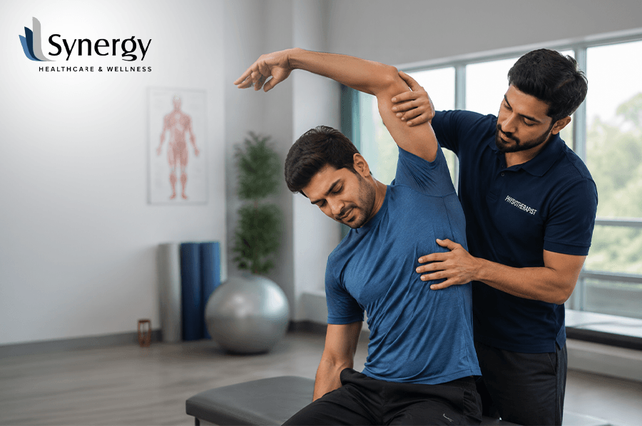
Pain that limits your movement, robs you of sleep, and affects your quality of life deserves more than just temporary relief. Manual therapy techniques are a clinically proven, hands-on approach to addressing the underlying causes of musculoskeletal pain, such as joint stiffness, soft tissue restriction, nerve irritation and postural dysfunction. This guide looks at the most successful techniques used in modern physiotherapy and why they always produce lasting results. When Pain Becomes a Daily Routine, and Why That Should Not Be the Case There’s a familiar pattern that emerges in people who have chronic pain. They rest and hope it gets better. When it does not, they reach for over-the-counter pain relief. They stretch it out a bit, maybe ice it and wait for the pain to go away on its own. Sometimes, it does. Sometimes it does not. What tends to linger, in the shoulder that aches through every night, in the lower back that tightens within an hour of sitting, in the knee that clicks and swells after a short walk, is pain that has been masked rather than dealt with. The root cause is left completely unaddressed. It is precisely in this situation that manual therapy techniques modify the clinical picture. Instead of numbing the pain signals, skilled manual therapy finds and treats the physical dysfunction that is sending the signals, whether it is a restricted joint, a pinched neural structure, a shortened fascial layer, or an overworked group of soft tissues compensating for a structural problem somewhere else. There is no small difference between managing pain and treating the cause. It’s the difference between short-term relief and real, lasting recovery. What Manual Therapy Really Is And Is Not? Manual therapy is a special area of physiotherapy practice in which skilled, hands-on clinical techniques are applied to joints, muscles, nerves and connective tissue. It is delivered by trained physiotherapists who use detailed anatomical knowledge and refined tactile assessment skills to identify and treat dysfunction that is often invisible on imaging. Soft tissue techniques have some superficial similarities, but it is not massage in the general sense. It’s not manipulation without clinical assessment. And it is not a single technique. It is a clinical framework comprising many different approaches, each with its own specific indications, mechanisms, and evidence base. Manual therapy is a central clinical role in non-invasive treatment strategies for musculoskeletal pain, providing individualised interventions that target biomechanical dysfunction and pain, with individualised assessment remaining the cornerstone of safe and effective practice. nih The Most Clinically Effective Manual Therapy Techniques 1. Joint Mobilisation Joint mobilisation is the use of carefully graded, rhythmic movements to a specific joint to restore its normal range of motion, reduce pain and improve mechanical function. Joint mobilisation is one of the most commonly used and best researched Joint mobilisation techniques in physiotherapy practice. This technique ranges in grade from gentle oscillatory movements (mainly for pain relief) to larger amplitude movements at end range (to restore mobility). It is often employed to: Studies have demonstrated that joint mobilisation can improve activation of deep stabilising muscles such as the lumbar multifidus and transverse abdominis, confirming that the benefits of this treatment extend beyond immediate pain relief, to include significant improvements in the underlying muscular support and spinal stability. 2. Release of Myofascia Fascia is a network of connective tissue that surrounds and links the body’s muscles, bones, nerves, and organs together. When this tissue becomes restricted through injury, repetitive loading, or prolonged posture, it develops tension patterns that develop pain, movement restrictions, and changes in biomechanics in several areas at the same time. Myofascial Release is a technique that involves applying sustained pressure to the restricted fascial layers to assist the tissue in lengthening and releasing. Myofascial release involves the application of specifically directed, low load, long duration mechanical forces to the myofascial complex to bring about optimal tissue length, pain reduction, and improved function and, when used alongside conventional treatment, is effective in providing immediate pain relief and tissue tenderness. clinicaltrials.gov A systematic review and meta analysis of randomised controlled trials also showed that the longer the myofascial release intervention, the significantly better the outcomes for joint range of motion, especially at the cervical spine, underlining the benefit of ongoing, progressive treatment over time. 3. High-Velocity Low-Amplitude (HVLA) Thrust Manipulation HVLA manipulation is a high velocity, low amplitude thrust delivered in a specific direction to a specific spinal or peripheral joint. This is the procedure most commonly related to the audible “click” or “pop” reported by many patients. This technique is particularly effective in: A comprehensive review of randomised controlled trials, systematic reviews and meta-analyses published between 2003 and 2024 found that the Kaltenborn-Evjenth mobilisation method and HVLA manipulation both showed notable effectiveness in improving range of motion and reducing chronic pain, particularly in patients with segmental joint hypomobility. HVLA is not appropriate for all patients and needs appropriate clinical screening before application. When properly indicated and skilfully done, it often results in rapid and marked improvement in pain and mobility. 4. Techniques for Soft Tissue Soft tissue manual therapy includes a range of hands-on methods focused on treating muscles, tendons, ligaments, and the surrounding connective tissues. Each of these techniques targets a different level of soft tissue dysfunction and is chosen according to the specific findings of the clinical assessment. 5. Mobilisation With Movement (MWM) – Mulligan Concept The Mulligan Concept is the use of sustained joint accessory glides by the therapist with simultaneous active movement by the patient. The combined approach often provides immediate gains in pain free range of motion, often within a single session. It is particularly useful for peripheral joint conditions such as ankle sprains, knee stiffness, shoulder dysfunction and lateral epicondylalgia. How Do Manual Therapy Techniques Work In the Body? Patients learn more about these techniques and how they work, and become more involved in their treatment. Mechanism Efficacy Cavitation Transient decrease in intra articular pressure; suppression of pain through neurological pathways Fascial lengthening Decreased tissue
Physiotherapy for Sedentary Lifestyle and Long Sitting Effects
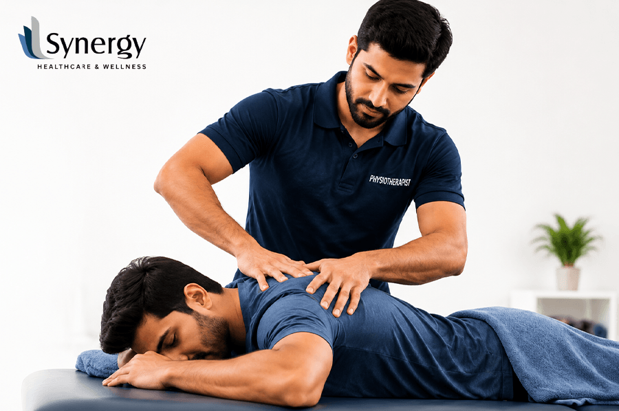
Spending hours at a desk each day does far more damage than most people realise. The toll of a sedentary lifestyle is cumulative and subtle, ranging from chronic back pain and weak muscles to bad posture and limited joint mobility. Physiotherapy for sedentary lifestyle rovides a structured, evidence based route to undoing this damage, restoring movement, reducing pain, and rebuilding the physical resilience that prolonged sitting steadily erodes. When Sitting All Day Feels Like a Full-Body Problem You end a long day at work and stand up only to feel stiffness shoot through your lower back, tightness grip your hips, and a dull ache settle across your shoulders. You stretch for a few seconds. It helps briefly. You sit back down. And tomorrow, the same thing happens again. For millions of working adults, this cycle feels normal. It has been happening long enough that they have stopped questioning it. The back pain is just part of the job. The neck tension is just stress. The fatigue at the end of the day is just life. But none of these things is simply part of life. They are consistent physical signals that the human body is responding poorly to the demands being placed on it. Long hours of sitting, repeated across months and years, produce real and measurable changes in the body. Understanding what those changes are and why physiotherapy for sedentary lifestyle is one of the most effective ways to address them begins with understanding what sitting actually does to your physiology. What Prolonged Sitting Actually Does to the Body? The human body is not designed for extended stillness. Every system, muscular, skeletal, circulatory, neurological functions better with regular movement. When that movement is removed for hours at a time, day after day, the consequences accumulate across multiple areas simultaneously. Musculoskeletal Effects of Long Sitting Research confirms that prolonged sitting places a significant force on the muscles, increasing the risk of injury. Studies consistently show that people with desk-based jobs develop tight iliopsoas muscles, resulting in reduced strength, restricted hip movement, increased pelvic tilt, and excessive curvature of the lower back. These changes do not happen suddenly. They develop gradually so gradually that most people adapt to the discomfort without realising how significantly their movement patterns have changed. Common musculoskeletal consequences of a sedentary lifestyle include: Cardiovascular and Metabolic Consequences The physical damage extends beyond muscles and joints. Physical inactivity is recognised as the fourth leading risk factor for global mortality, accounting for six per cent of deaths worldwide. Research indicates that individuals who engage in prolonged sedentary behaviour, particularly alongside low levels of physical activity, face significantly elevated risks of cardiovascular disease and metabolic dysfunction. Evidence further suggests that persons who participate in long periods of sedentary behaviour, particularly in combination with low physical activity levels, are at high risk of reduced cardiorespiratory fitness and are therefore at greater risk for cardiovascular mortality. Cognitive and Fatigue Related Effects It is not only the body that suffers. Studies suggest that spending extended periods sitting is linked to increased general and physical fatigue, along with lower overall activity levels among the general population. Concentration dips. Decision making slows. Productivity, the very thing most desk workers are trying to protect, is quietly undermined by the environment built around maximising it. How Physiotherapy for Sedentary Lifestyle Reverses the Damage? Area of Concern Physiotherapy Approach Expected Outcome Lower back pain Core strengthening, spinal mobilisation Reduced pain, improved stability Hip flexor tightness Targeted stretching, muscle release Restored hip mobility Poor posture Postural re-education, ergonomic advice Corrected alignment Weak gluteal muscles Progressive resistance training Better pelvic support Neck and shoulder tension Manual therapy, mobility exercises Reduced tension headaches Reduced cardiovascular fitness Graded aerobic exercise programme Improved endurance Sedentary lifestyle physiotherapy is not a single treatment. It is a structured, personalised programme that addresses the specific impairments each individual has developed based on their lifestyle, posture, and daily demands. Assessment First and Treatment Second A thorough physiotherapy assessment identifies which muscles have shortened, which have weakened, where movement has become restricted, and how posture has adapted over time. This assessment prevents generic treatment and ensures that every exercise and technique used targets the actual cause of discomfort rather than simply managing symptoms. The Core Components of Effective Sedentary Lifestyle Physiotherapy Postural Re-Education: Most people with desk-based jobs do not know what neutral spinal alignment actually feels like in their bodies. Physiotherapy teaches this not as an abstract concept but as a felt, repeatable physical experience that can be applied during working hours. Targeted Strengthening: The muscles most affected by prolonged sitting, the deep core, the gluteals, the thoracic stabilisers, require progressive strengthening to restore their function. This is not general gym work. It is specific, clinically directed exercise that addresses precise muscular deficits identified during assessment. Flexibility and Work Mobility: Tight hip flexors, stiff thoracic spines and restricted shoulder complexes all require specific flexibility work. Active stretching can be complemented with manual therapy techniques such as joint mobilisation and soft tissue release to restore range of motion more efficiently than stretching alone. Ergonomic Guidance: Sedentary lifestyle physiotherapy extends beyond the clinic. Advice on workstation setup, screen height, chair positioning, keyboard placement, and break frequency, ensures that the improvements made during treatment are not undone by the same environment that caused the problem in the first place. Movement Habit Development: Research suggests that an increase in daily walking steps is positively associated with improvements in fitness, making structured movement throughout the day a clinically relevant strategy for those reducing sedentary time. Physiotherapists provide specific guidance on how to integrate effective movement breaks into a working routine without disrupting productivity. For further guidance on exercises that complement your in-clinic physiotherapy programme, explore Synergy Physio’s home exercise resource, a practical reference for building daily movement habits between sessions. Practical Steps You Can Take Today Whilst professional physiotherapy assessment provides the most accurate and personalised guidance, several evidence informed habits can support recovery and reduce ongoing damage: For further evidence based
Why Evidence Based Physiotherapy Ensures Better Recovery Results
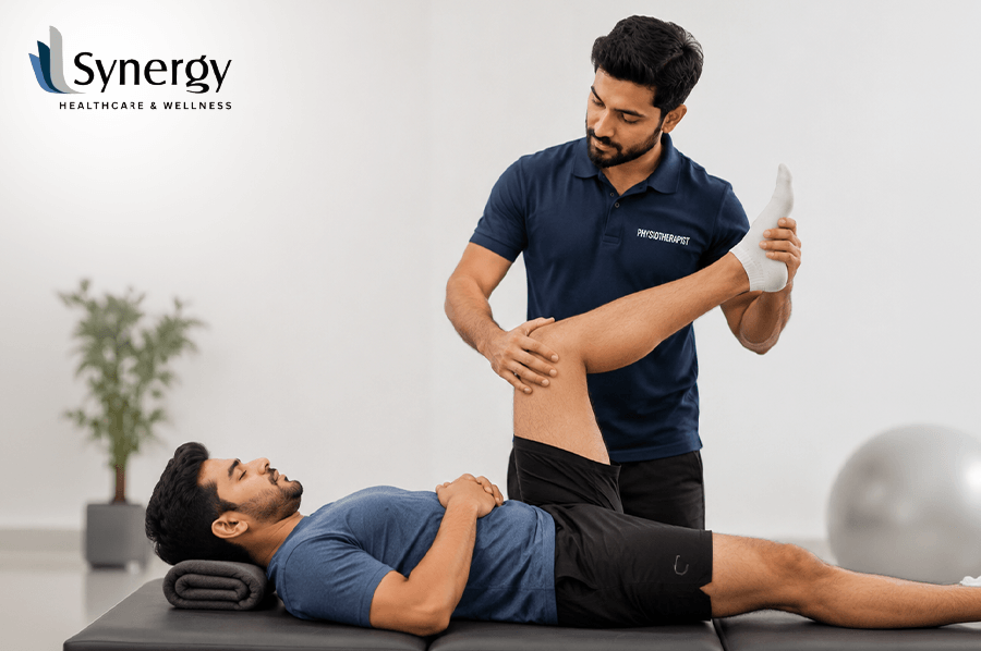
Recovery from injury, surgery or neurological conditions is seldom a simple process. Evidence based physiotherapy removes the guesswork from treatment, with all decisions based on clinical research and measurable results. This approach delivers quicker, safer and more reliable results in recovery and is considered the gold standard in modern rehabilitation care, from post surgical rehabilitation to gait training physiotherapy. What Happens When Recovery Stalls, and How You’re Treated Your sessions are finished. You do the exercises. Weeks go by. But something still does not feel right. The pain is there, the movement does not return as hoped, or progress simply stalls for no apparent reason. Most people don’t understand how common this experience is. In many cases, it’s not a matter of effort. It’s a question of method. Physiotherapy is not a uniform practice. The techniques used, the course of treatment, and the instruments used all vary greatly depending on the practitioner or clinic. Often, the framework that guides clinical decisions is what separates a recovery that moves forward steadily from one that stalls. When done right, that framework is evidence based physiotherapy. Understanding Evidence Based Physiotherapy and why it gets better outcomes Clinical evidence shows physiotherapy is the systematic application of the best available clinical research, together with the therapist’s professional expertise and the individual needs of the patient, to guide all aspects of treatment. It is not one technique. It’s a clinical philosophy. One that asks a specific question at every decision point: What does the research show works best for this condition, in this patient, at this stage of recovery? The 3 Pillars of Evidence Based Practice Pilar Description Best Available Science Evidence from peer-reviewed clinical trials, systematic reviews and meta-analyses Clinical Expertise The trained judgement, assessment skills and professional experience of the therapist Patient Values and Goals The individual’s interests, lifestyle and goals for recovery When all three pillars are working together, treatment is much more accurate. We don’t prescribe exercises because they are routine; we prescribe them because the evidence shows they are effective for a specific condition. No random event, but an evolution powered by clinical markers that can be measured. This strategy reduces the chance of a prolonged recovery, prevents excessive stress on injured tissue, and ensures patients don’t get outdated treatments that have been replaced by better ones. Evidence from peer-reviewed rehabilitation literature suggests that patients receiving evidence-informed physiotherapy programmes have significantly better functional outcomes than those receiving generic or protocol driven care. How Physiotherapy for Gait Training Can Enhance Walking Recovery? Gait training is one of the most compelling examples of clinical evidence physiotherapy in action. It is a structured, research led approach to restoring normal walking patterns in patients recovering from stroke, surgery, orthopaedic injury or neurological conditions. What Gait Training Really Means? Gait training physiotherapy is much more than helping someone to walk again. It involves a detailed assessment of how you move, the sequence in which your muscles activate, how you maintain balance, and how effectively you can move around. This is followed by a progressive programme tailored to address the specific issues that are limiting your recovery. Common techniques used in evidence based gait training are: The Evidence for High-Intensity Gait Training Growing clinical evidence supports higher intensity gait training in structured physiotherapy programmes. Research published in 2025 confirms that high-intensity cardiovascular gait training with a focus on stepping practice improves gait function after stroke and appears to be superior to standard approaches at lower intensity. This is also supported by studies that have found that intensive and accurate training supports neuronal plasticity and motor recovery, especially when combined with traditional physiotherapy approaches. This body of evidence contributes to a crucial point: recovery from walking is not a matter of time. It’s the right stimulus at the right intensity, coupled with clinical reasoning that is research-based. Physiotherapy gait training is a structured, evidence-based pathway back to confident, independent movement for patients recovering from stroke, knee replacement, spinal injury or prolonged immobility. The Dangers of Non Evidence Based Treatment Knowing what happens when physiotherapy is untethered from evidence is useful. Without a research informed framework, treatment can become a matter of habit, not effectiveness. Exercises are prescribed based on tradition and not on clinical outcome data. Progressions can be too slow, which delays recovery unnecessarily, or they can be too aggressive, risking reinjury. Furthermore, passive interventions, such as heat packs or basic massage, can be given as the main intervention when evidence clearly shows active rehabilitation to be the superior long-term strategy. Sometimes, non evidence based care does not cause harm. But it is rarely that efficient. And rehab, there’s no time to lose. Every week of delay in recovery has a real physical, emotional and in many cases financial cost. Why Choose Evidence-led rehabilitation: Key Benefits Synergy Using Research driven physiotherapy to Guide Every Treatment Plan At Synergy, the commitment to evidence based physiotherapy is not a marketing line. It is the clinical basis on which all assessment, diagnosis and treatment programmes are built. Synergy has clinics across Chennai in Anna Nagar, T. Nagar, Adyar, Mogappair and Vepery. We integrate the latest clinical research and our deep knowledge of therapy to provide rehabilitation that is precise, measurable and most importantly, effective. If you’re recovering from a sports injury, managing chronic pain or regaining your walking ability with gait training physiotherapy, Synergy’s team applies the same rigorous approach to your care: assess thoroughly, treat specifically, and progress systematically. This builds a process of recovery that is transparent, traceable, and evidence-based of what really works. Discover the Synergy approach to physiotherapy exercises for home-based recovery, a practical extension of your in-clinic treatment plan. Conclusion: Restoring Movement with Science, Skill, and Care Recovery is seldom a straight line. There are good days, slow days and times when the progress seems invisible. The difference that keeps progress moving in the right direction is the quality of the clinical thinking behind your treatment. Evidence based physiotherapy makes the process less uncertain.