
Tennis & Golfers Elbow: Symptoms, Causes & Treatment
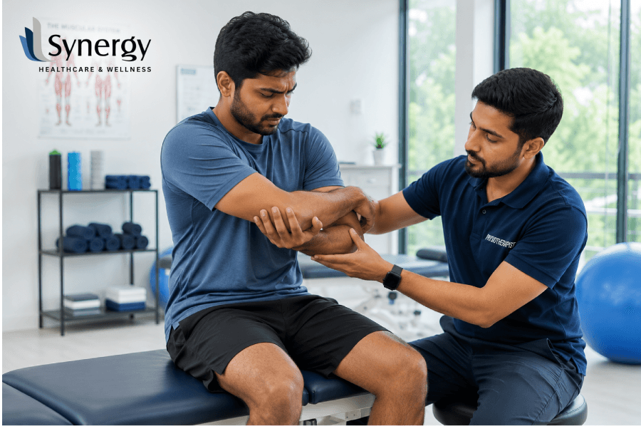
Epicondylitis is a common source of chronic forearm pain. Tennis elbow is an injury to the outside of the elbow tendon, and golfer’s elbow is an injury to the inside of the elbow tendon. Treatment for this structural strain is targeted rest and specific loading exercises, together with evidence-based clinical rehabilitation. This blog outlines the symptoms, biomechanical causes and clinical interventions, including home elbow pain treatment at home to restore painless upper limb mobility. Intro: Hidden Causes Behind Tennis and Golfers Elbow in Daily Life Repetitive forearm rotation, heavy lifting and poor wrist mechanics place excessive stress on the tendons that attach to the elbow joint. These injuries are medically known as lateral epicondylitis (tennis elbow) and medial epicondylitis (golfers elbow). The names sound athletic, but the conditions are experienced mostly by people doing routine manual labour, office work or repetitive industrial tasks. The key to selecting the right path to recovery is understanding how these conditions come about. Many mild or early-stage tendon strains can be treated at home with targeted elbow pain treatment at home. Chronic or serious cases require professional evaluation to avoid permanent degeneration of tissue. Tennis Elbow vs. Golfer’s Elbow: What’s the Difference? In order to properly treat upper limb pain, you need to first identify what tendon structures are involved. The elbow joint is the point of attachment for the muscles that move the wrist and fingers. These are the dissimilarities in the structure of these two diseases: Aspect Tennis Elbow (Lateral Epicondylitis) Golfer’s Elbow (Medial Epicondylitis) Primary Area (Located Anatomically) Lateral epicondyle (Outer part of the elbow). Medial epicond (Inner part of the elbow). Group of Affected Tendons Extensor carpi radialis (Common extensor tendon). Flexor carpi ulnaris (Wrist flexors and pronators). Aggravating Movements Lifting objects with palms down, typing, and turning doorknobs. Gripping tight, wrist bending, crushing and throwing. Radiation pathway of pain Radiates down the back of the forearm into the wrist. Radiates down inside the forearm towards the ring finger and pinky. Key Symptoms and Red-Flag Signs The recognition of early signs of epicondylitis prevents acute tendon irritation from evolving into chronic tendinopathy. Both conditions cause localised joint soreness, but their patterns of diagnosis are different. Symptoms of Tennis Elbow Golfers Elbow Symptoms Daily Biomechanical Triggers & Root Causes Tendons are much less vascularised than muscles, and therefore very prone to micro-tearing when subjected to sudden overload or repetitive mechanical stress. Evidence-Based Self-Care for Successful Treatment of Elbow Pain at Home In mild cases, you can reduce inflammation and start tissue repair by following a structured routine of elbow pain treatment at home. 1. The POLICE Protocol Modern sports medicine has moved away from absolute rest and recommends the POLICE protocol for early tendon management: 2. Exercises To Be Done At Home Following the resolution of pain, progressive overload is used to recondition the tendons: Treatment Procedures Are you dealing with pain in multiple locations? Do you need help with your chronic pain? Read more about how we use advanced treatments for chronic pain to recover naturally through physiotherapy. Synergy Healthcare Comprehensive Care If home management does not provide long-term relief, then clinical intervention is required to address deep tissue degeneration. Synergy Healthcare’s senior consultants provide a high level of evidence-based physiotherapy specific to your biomechanical needs. Instead of just covering up pain, clinical protocols aim at structural alignment, tissue regeneration and long-term joint health. Our clinical teams in Anna Nagar, T. Nagar, Adyar, Mogappair and Vepery use advanced diagnostic techniques to distinguish between simple tendon strain, nerve entrapment and ligament laxity. We determine the precise time of injury and tailor rehabilitation exercises that ensure permanent effects. Final Thoughts on Dealing with Joint Strain Treating ongoing upper limb discomfort requires a combination of proactive home elbow pain treatment at home and expert clinical advice. If you disregard early symptoms like a weak grip or tenderness in the inner joint, you can develop chronic tendinopathy from a simple strain. You’ll have a definitive diagnosis and a clear plan for recovery when you work with the expert team at Synergy. Acting quickly preserves the health of your tendons, regains your physical strength and allows you to continue comfortably in your daily life. Take control of your recovery today with expert guidance from Synergy and restore your strength with confidence and precision. FAQs 1. How long does golfers elbow take to heal completely with home care? Symptoms generally subside within 4 to 6 weeks with home care and rest, but chronic tendon problems can take several months to fully recover, with structured physical therapy. Getting some early rest and loading is important for faster tendon healing. 2. If I am diagnosed with tennis elbow, can I keep weight lifting? During the acute painful phase, no heavy lifting should be performed, as pushing through the sharp pain can exacerbate the micro-tears in the tendon and increase overall recovery time. Normal, safer movements are modified or pain-free in the early stages of healing. 3. Why do I have pain on the inside of my elbow when I am not playing golf? Everyday activities like lifting bags, typing or using poor wrist alignment place the same mechanical stress on inner elbow tendons as a golf swing. More often than not, repetitive daily strain contributes more than sports activity alone. 4. Do I use a heating pad or an ice pack for acute tendon pain? Generally, ice packs are recommended for 48 to 72 hours to help reduce acute inflammation. Heat pads are more useful later, to relax stiff muscles before stretching. Alternating methods may have the right intensity for an optimal enhanced recovery. 5. Will the counterforce brace be effective against epicondylitis? The purpose of the counterforce brace is to take off load from the tendon insertion point and spread out mechanical loads across the arm. The counterforce brace will be most effective in combination with rest and exercise. 6. What will happen if I neglect the pain in my forearm tendon? Neglecting this condition may
Calf & Leg Muscle Pain: Causes, Treatment & Exercises
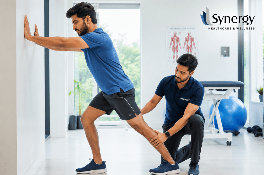
Persistent pain in the calf muscle pain causes in your leg can really affect your mobility, sleep, and quality of life. This clinical guide covers the main calf muscle pain causes, from simple dehydration to acute injuries of the gastrocnemius muscle pain pathway. Read on for professional calf pain treatment options, targeted home interventions and the ideal exercise for calf muscle pain to get long-term calf pain relief. Lower Limb Discomfort: An Introduction Every day, thousands of people, from professional athletes to desk-bound corporate workers, suffer from lower limb problems, particularly pain in the back of the lower leg. The calf is a complex arrangement of muscles, tendons, nerves and blood vessels that are constantly working to keep the body upright, absorb impact from the ground and push the body forward as you walk. When the distress is localised, the first step to sustainable calf muscle pain relief is to identify the exact triggers. The final course of recovery depends on whether the discomfort is due to lifestyle fatigue, an acute mechanical tear, or an underlying circulatory problem. What Makes the Lower Leg Hurt? To treat problems in the lower limb, we need to assess the common physiological and anatomical factors that interfere with normal muscular function. The calf is very susceptible to mechanical overload, biochemical changes and vascular changes. 1. Acute Muscle Strains & Mechanical Muscle Tears A common reason for sudden, sharp pain is a muscle strain. This happens when the structural fibres are overstretched or torn by explosive acceleration, sudden changes of direction or poor warm-ups. During competitive sports or sudden physical exertions, the calf’s lateral and medial bellies are particularly vulnerable. 2. Night cramps and involuntary spasms Their sudden, intense contraction in the night often wakes people up from deep sleep. These involuntary spasms are closely associated with localised muscle fatigue built up during daytime standing, minor electrolyte imbalances (such as low magnesium, potassium or calcium) or mild dehydration. 3. Tendon Overuse (Chronic) The Achilles tendon connects the main muscles of the calf to the heel bone. Achilles tendinopathy is a condition which can be caused by repetitive micro-trauma from running on hard surfaces, inappropriate footwear or structural biomechanical abnormalities. It is characterised by chronic stiffness and burning sensations in the lower leg. 4. Failures of Circulatory and Vascular Systems A decreased return of blood from the lower extremities can cause deep, throbbing aches. Some other conditions, like chronic venous insufficiency or deep vein thrombosis (DVT), carry significant clinical risks and require urgent medical evaluation instead of routine physical rehabilitation. Type of Cause Initial Symptom Presentation Commonly Aggravating Factors Suggested first approach Acute Sprain Sharp, stabbing pain. Localised swelling, Bruising. Sudden acceleration, bouncing off Rest, ice, clinical evaluation. Cramp at Night Shooting, transient locking spasm; postcramp ache Standing still for a long time, dehydration Light static stretching, hydration management Tendonopathy Morning stiffness; burning sensation in the heel Unsupportive shoes, running over and over Graded tendon loading, eccentric exercise Gastrocnemius Muscle Pain: Clinical Perspective The calf complex consists of two important muscles, the deeper soleus and the superficial, double-headed gastrocnemius muscle. The gastrocnemius crosses 2 major joints (the knee and ankle ), so it is very active during powerful lower limb movement. Gastrocnemius muscle pain is usually caused by a sudden eccentric contraction; for example, lunging forward in tennis or sprinting to catch a bus. This strain usually manifests as a distinct “popping” or snapping sensation in the back of the leg, followed immediately by swelling, local tenderness, and an inability to perform a standard heel-raise. This presentation requires management with a structured clinical approach so that the structural collagen fibres heal in optimal alignment and do not result in chronic tightness or recurrent tears. Total Solutions for Lower Leg Care To effectively relieve leg calf muscle pain, two strategies are needed: controlling acute symptoms and proactive physical rehabilitation. Immediate Home Care: The R.I.C.E. (Modified) Protocol In the case of serious structural problems, the classic conservation framework of the first 48 hours can speed up recovery: Clinical Modalities & Specific Interventions Where home therapy fails to provide relief from the painful calf muscle, professional physical therapy presents advanced techniques. Targeted manual therapy releases structural myofascial trigger points, localised ultrasound increases circulation in deep tissues, and therapeutic taping reduces mechanical strain on healing fibres, say clinical specialists. Benefits of Active Rehabilitation Progressive movement is one of the best healing tools for soft tissue. Proper stretching in combination with strengthening exercises will help to heal soft tissues and gain strength. 1. The Classic Wall Assisted Calf Stretch This movement restores ideal length to shortened muscle fibres and provides quick relief from calf pain during or following a tight episode. Stand facing a solid wall. Extend both hands in front of you at shoulder level to balance yourself. Step back with the painful leg, keeping the heel firmly pressed into the floor, the rear knee straight. Bend your front knee gently and lean your torso forward until you feel a deep and comfortable pull through the back of your lower leg. Hold steady for 30 seconds without bouncing and repeat the sequence 3 times. 2. Sitting Towel Mobilisation Sit on a flat surface with your lower limbs stretched directly ahead. Loop a long non-elastic exercise band or a clean bath towel securely around the ball of your foot. Hold tight to both ends of the towel and pull it slowly backwards towards your body, bringing your toes up towards your shins. The exercise works to relieve calf pain without placing pressure on an injured leg. Maintain tension for 25 seconds. Breathe slowly and then relax. 3. Double Heel Raises Once the acute irritation has passed, it is important to build up the tissue capacity to avoid future strains. Stand flat on the floor near a counter or wall for balance support. Slowly lift both heels, pressing your body weight up onto the balls of your feet over a count of three seconds. Hold at the top of the contraction,
Foot Pain: Why It Happens and How to Get Relief at Home
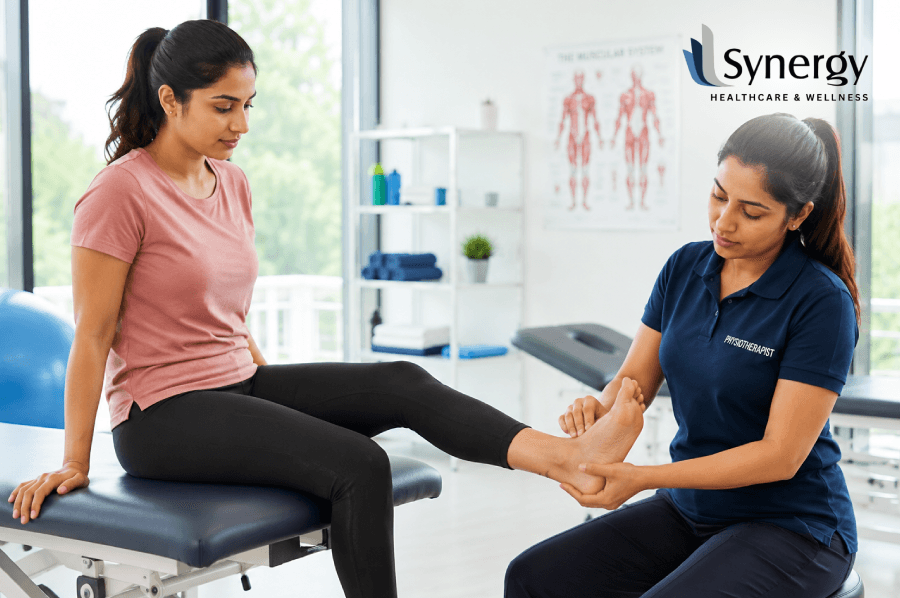
A lingering ache or pain under foot can greatly impact your daily life, making simple mobility a stressful challenge. This comprehensive guide explores the underlying structural reasons for discomfort in the lower extremities, including plantar fasciitis and mechanical strains, and offers effective, evidence-based home remedies for feet pain to help you regain comfort, realign structure and walk pain-free. Intro: Why does Every Step feel like you are walking on a bruise? Walking is made possible by the complex and well-coordinated interaction of the body’s bones, muscles, ligaments, and tendons. Disturbance of this fine mechanical network will frequently result in a nagging, sharp, or throbbing pain under foot. Whether you are experiencing an annoying pain in sole of foot after long hours of standing or a deep, aching pain in middle of foot, understanding the biomechanics of your lower limbs is the critical first step toward long-term rehabilitation. Even though temporary discomfort may appear to be a minor inconvenience, ignoring persistent foot instep pain or structural soreness can change your natural gait, causing secondary problems in your ankles, knees, and lower back. Knowing the root causes of your pain allows you to utilise specific non-invasive recovery methods and potent home remedies for aching feet from the privacy of your living room. Other Causes of Arch and Sole Discomfort The trick to getting relief from lower limb pain is to be very precise about where the irritation is coming from. Sudden changes in physical exertion, unsuitable shoes, and biomechanical irregularities may place excessive stress on the lower extremities. Exact Location of Pain Most Frequent Underlying Clinical Cause Major Structural Symptoms Pain in sole of foot (near the heel) Fasciitis (plantar) Very first steps in the morning, sharp stabbing pain. Discomfort located in the arch of the foot Over-pronation or Midtarsal Joint Stress Deep, dull pain along the inner arch is worse with weight-bearing. Side of foot (Lateral margin) Jones Fracture or Peroneus Tendonitis Sensitivity to pressure, localized edema, and intermittent and sharp discomfort when walking over rough ground. Pain in lower feet / Metatarsals / Feet Metatarsalgia or Morton’s Neuroma By a sharp pebble, or walked on it. The pain of the acute foot that occurs in people after walking is usually an inflammatory warning sign that the soft tissues have been overloaded beyond their physiological capacity. This is especially so for those with foot instep pain, which directly affects the structural integrity of the medial longitudinal arch. Small structural micro-tears can become chronic and debilitating with the right mechanical rest and tissue care. Best Home Remedies for Foot Pain and Physical Care You can treat lower extremity discomfort at home without spending a fortune on equipment. A systematic, organised routine can do wonders for localised inflammation, irritated nerve pathways and can speed up the natural soft-tissue healing process. 1. Rest, Ice, Compression and Elevation (R.I.C.E.) The classic R.I.C.E. protocol is the basic feet pain relief solution to acute structural inflammation. Rest Avoid high-impact activities that aggravate the side of foot or arch strain. Ice Apply an ice pack covered with a cloth to the area for 15 to 20 minutes, three times a day. Rolling the sole of the foot over a frozen water bottle is an excellent targeted deep tissue massage that cools inflamed fascia. Compression & Elevation Use a supportive elastic bandage to reduce localised swelling. Elevate your lower limbs on pillows above heart level to encourage healthy venous return. 2. Targeted Stretching for Plantar and Calf Tightness of the gastrocnemius and soleus (calf muscles) directly increases the mechanical strain on the lower foot structures. Gentle controlled calf stretches against a wall twice daily will help to decrease pain in middle of foot structures. Also, before you get out of bed, gently pull your toes back toward your shin for 30 seconds to pre-stretch the plantar tissues and avoid sharp morning twinges. 3. Tennis Ball Self-Myofascial Release To relieve general foot pain, sit down and place a tennis ball or a dedicated reflexology roller underneath the arch of your foot. Apply moderate but comfortable downward pressure and roll the ball slowly from the base of your toes to your heel. This simple practice breaks up painful soft-tissue adhesions, increases localised blood circulation, and alleviates persistent foot pain after walking a strenuous day of movement. When to Seek Urgent Medical Advice? Looking for the best painkiller for foot pain – over-the-counter non-steroidal anti-inflammatory drugs (NSAIDs) can temporarily take the edge off severe discomfort, but medication should never be used as a long-term cure. If you have an absolute inability to bear weight on the pain in lower feet, visible structural deformities or progressive numbness, you must seek immediate medical advice from a qualified healthcare provider. For further information on the clinical benefits of professional therapeutic intervention on tissue repair at all stages of the structural healing process, you can view the full medical summary offered by the Chartered Society of Physiotherapy or the comprehensive lower limb clinical guidelines hosted on Arthritis Research UK. Synergy Professional Solutions in Biomechanics While focused home remedies for feet pain may be great for quick symptom relief, actual structural healing needs a more thoughtful assessment of your overall movement patterns. Temporary fixes often cover up the real mechanical issues, such as poor foot position, muscular imbalances, or an uneven gait. Long-term, if you sense that the discomfort in your structure is not improving with routine rest, then getting an objective, evidence-based physical assessment is essential to preserving your long-term mobility. At Synergy, our clinical team is trained to find the true cause of your physical discomfort, rather than just treating the symptoms on the surface. With highly advanced diagnostic tools, including detailed digital gait analysis, our senior rehabilitation experts can track exactly how weight is distributed across your lower limbs with each step you take. This exact, data-driven approach is extremely helpful for those with chronic foot pain after walking, because it reveals subtle structural imbalances that may be stressing the soft tissue. Whether
Middle & Lower Back Pain: Causes, Red Flags & Relief
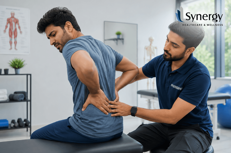
If you are dealing with consistent spinal tightness or unexpected pain, it’s important to understand the root causes behind your symptoms. This guide explores the mechanical factors causing middle back pain and spinal imbalance of the lower back area, examines significant warning signs which must be acted upon urgently, and provides a rehabilitation protocol based on scientific principles for restoring good posture and mobility. Introduction: Why Is Your Middle Back Sending You Warning Signals? The spine in the human body is a complex load-bearing structure that helps coordinate our daily movements, balance and structural integrity. Discomfort can occur in different areas when structural imbalances occur. While a great deal of clinical attention is focused on lumbar issues, middle back pain is an increasingly common issue that impacts office workers, athletes and older adults alike. Mid back spine pain discomfort may be a dull, constant ache or a sharp, constrictive spasm that limits deep breathing and rotational movement. In order to ensure musculoskeletal wellness in the long term, it is crucial to understand the cause, identify whether there is any benign muscular strain or not, and know when to get medical help. Postural Strain & Mechanical Cause The middle back contains 12 vertebrae (T1-T12). These vertebrae are tightly attached to your rib cage. This structure provides essential stability and protection to the internal thoracic organs. But the common sedentary lifestyle today often puts this area under static load and repetitive stress. Sitting in badly designed office chairs, staring at mobile phones or leaning forward over laptops for extended periods causes the shoulders to roll forward and leads to a hunched posture. This forward head carriage stretches the posterior thoracic ligaments over time and causes the rhomboids, trapezius and erector spinae muscles to work excessively to keep the torso upright. This chronic overworking of the muscles results in ongoing fatigue, localised inflammation and deep, aching discomfort across the mid back spine pain area. Timeline Postural Discomfort Stage 1 Sometimes get stiff after long hours at the desk, but it goes away with movement. Stage 2 Dull aches repeated for hours. Muscle tension evident. Stage 3 Sharp, throbbing pain that limits your ability to turn and take deep breaths. Stage 4 Chronic daily limitation, severe tightness in tissues and nerve tension. Traumatic And Degenerative Causes In addition to the usual postural imbalances, there are a number of structural abnormalities and mechanical problems that can cause chronic back pain: Muscle Strains and Ligament Sprains The supportive soft tissues of the middle and lower back can be subject to micro-tears when sudden twisting movements, improper heavy lifting or unaccustomed physical exertion occur. Thoracic Disc Herniations Because the rib cage is so rigid, disc herniations in the thoracic spine are relatively uncommon. When they do happen, they can compress the intercostal nerves and cause pain to radiate around the rib cage. Vertebral Compression Fractures Small hairline fractures can occur in the vertebral bodies of older adults, often due to underlying systemic conditions like osteoporosis, and cause a sudden onset of severe middle back pain. Osteoarthritis and Spondylosis Degenerative changes in the facet joints of the spine with age reduce the shock-absorbing function of the intervertebral discs. This causes stiffness in the morning and mechanical friction between joints. Spinal Dynamics: Thoracic vs. Lumbar Systems When devising an effective management strategy, it is useful to compare the effect of structural stress on the thoracic spine to that on the lower lumbar and pelvic regions. The lower back has different structural demands than the mid spine, and therefore injuries of these areas have different clinical patterns: Area of the Spine Primary structural function Typical Pathological Findings Core Rehab Emphasis Thoracic Spine (Mid Back) Stability, protection of the internal organs, and support of the rib cage. Strain from posture, muscle fatigue, irritation of intercostal nerves and limited rotation. Scape retraction, thoracic extension mobility, and ergonomic modifications. Lower Back – Lumbar And Pelvic Spine Weight-bearing, shock absorption, multi-directional flexibility Disc bulges, sciatica, muscle spasms and local pelvic instability. Decompression of the lumbar, stabilisation of the core, and strengthening of the gluteal muscles. When considering lower back pain, clinicians often look closely at the pelvic foundation. The sacroiliac joints join the sacrum to the iliac bones of the pelvis. Dysfunction here results in distinct SI joint pain, which typically presents as a dull, unilateral ache in the lower back, buttocks, or upper thigh. This particular discomfort is frequently exacerbated by prolonged standing, climbing stairs, or asymmetrical weight-bearing activities. Important Clinical Warning Signs The vast majority of problems are mechanical and respond well to conservative care. There are clinical signs that need immediate medical evaluation. You need to watch out for these red flags to make sure there isn’t a systemic or structural complication: Unexplained Weight Loss or Fever If your back pain is accompanied by chills or a persistent fever, or if you have experienced unexplained weight loss, it could be indicative of an underlying systemic infection or oncological problem. Neurological Compromise Progressive numbness, pins-and-needles or weakness in the lower limbs needs urgent attention. Cauda Equina Syndrome is a medical emergency and is caused by compression of the lower spinal nerve roots. It presents with saddle anaesthesia and sudden loss of bowel or bladder control. Radiating Pain Pains that are sharp and occur along a route around the chest wall or running along the back of the leg (sciatica) indicate irritation of the nerve roots. Pain Waking You From Sleep You may find yourself awakening from deep sleep due to pain which is not relieved by slight adjustments in posture. This indicates a non-mechanical condition that needs evaluation. History of Trauma If your pain started after a fall, motor vehicle accident, or direct physical trauma, it is important to get an immediate medical evaluation to rule out structural fractures. Daily Tips for Long-Term Spine Health To have a strong, pain-free spine, it is important to build healthy habits that you can incorporate into your daily life. Small adjustments in the way
Guide to Neck and Shoulder Pain Relief
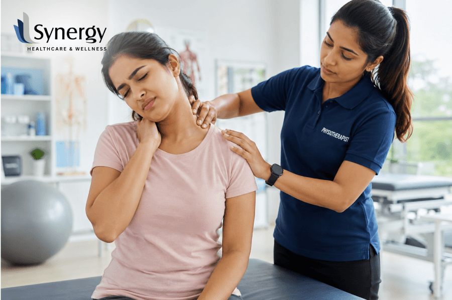
Chronic neck and shoulder pain can severely limit your daily life, making even small head movements painful. Supportive cervical collar devices provide immediate short-term relief through stabilisation of the cervical spine, but long-term recovery must be aimed at targeted rehabilitation. Knowing your options, postural corrections and when to see a specialist is important in order to achieve permanent pain-free, healthy mobility. Introduction: When Neck Pain Starts Controlling Your Routine and Focus You wake up in the morning, roll over to turn off your alarm and are immediately struck by a sharp, locking sensation that goes from the base of your skull straight down into your shoulder blade. Even simple daily activities, like checking your car’s blind spot while driving or looking down at your laptop screen, suddenly require a slow, guarded pivot of your entire upper body. That localised stiffness morphs into a heavy, burning ache by mid-afternoon, which sets off tension headaches and drains your focus. Instead of moving smoothly through your personal and professional routine, you are forever rubbing your neck, adjusting your desk chair, and frantically searching for a position that gives you a few moments of temporary physical relief. This draining physical cycle is a super common issue for office workers, active people, and older adults managing modern, screen-heavy lifestyles. Quick hot patches or over-the-counter anti-inflammatories can mask structural imbalances or age-related changes that settle into the upper spine, but the deeper problem remains. You must understand the exact physiological causes of your discomfort, the place of modern supportive devices such as a cervical collar, and dedicate yourself to a structured approach to spinal rehabilitation if a permanent breakthrough is to be made. Unravelling the Anatomical Root Causes of Sore Muscles Your human cervical spine is a very complex structure that carries the weight of your head while allowing movement in several directions. The muscles, ligaments and nerves in this area are closely linked so that any damage or inflammation in one area quickly spreads out, creating overlapping neck and shoulder pain. The first step to mapping out an effective treatment pathway is identifying exactly what is driving your symptoms. Ergonomic Stress and Chronic Postural Overload This is the modern “forward head posture,” often called tech-neck, and it is the most common cause of upper back tension. For every inch your head protrudes past the natural alignment of your spine, the relative weight on your lower cervical vertebrae increases by nearly five kilograms. This constant mechanical overload keeps the trapezius and levator scapulae muscles in a constant state of contraction, resulting in painful myofascial trigger points, decreased blood flow and chronic muscle fatigue. Structural Changes and Progressive Creep As we age, our spines go through a regular ageing process that slowly alters the structural integrity of the intervertebral discs and facet joints. When one goes for a consultation with a specialist, it is very important to know the real meaning of cervical spondylosis. This medical term refers to the progressive, age-related wear and tear affecting the cervical spine, characterised by the thinning of spinal discs, the development of protective bone spurs, and the gradual narrowing of the spinal canal. It usually causes some local stiffness of the joint and can sometimes pinch nearby nerve roots, causing radiating pain, numbness or pins and needles radiating down your arm to your fingers. The Role of Orthopaedic Supports for Acute Neck Trauma In case of acute injury to the upper spine, for example, in whiplash deceleration in a vehicle accident or in case of a severe flare-up of progressive degeneration in the joints, it is important to keep the movement to a minimum at once. A specialised cervical brace is a crucial first-line intervention that temporarily offloads mechanical stress, protects delicate nerve pathways and allows inflamed soft tissue to rest and recover. To guarantee a safe and effective recovery, it is important to become knowledgeable about the different types of cervical collars available in the medical market, as each design is engineered for a specific degree of structural restriction: Orthotic Type Main Material Degree of Mobility Impairment Best Clinical Usefulness Foam Collar Soft Breathable fabric knitted with high-density polyurethane foam Minimal restriction, mostly a tactile reminder to stay away from sudden rotations Mild muscle strains, early postural fatigue, and nighttime boundary support Semi-Rigid Neck Collar Moulded plastic borders with soft, built-in foam padding borders Moderate restriction. Restricts forces of flexion, extension and lateral bending. Severe nerve root compression, stable structural fractures, and acute whiplash injury Rigid Extrication Collar Two-piece hard polyethene shell with dedicated chin support ledge Maximum restriction: locks the upper cervical joints completely in the neutral position Critical post-surgical recovery, acute emergency transport, high-impact spinal trauma While a cervical collar for neck pain can provide immediate physical comfort in the initial stages of an injury, medical research stresses that these orthotic devices should be used selectively. Using a neck collar for neck pain beyond a few days without professional guidance can result in muscle atrophy and increased joint stiffness, which can ultimately delay your long-term recovery. Restoring Long-Term Mobility with Targeted Physical Rehabilitation A supportive brace will help a lot to get through an acute flare-up, but to get rid of upper spinal discomfort permanently, one must transition from passive immobilisation to active, movement-based therapy. To get back to full, pain-free movement, you need to re-educate your postural muscles, increase your joint flexibility and build up your deep structural endurance. The Power of Prescribed Exercise Therapy Customised physiotherapy exercises for neck pain are the best way to reverse chronic muscle tightness and protect degenerating spinal discs. These specialised routines are designed to activate the deep neck flexors, the internal core muscles of your neck, while stretching shortened chest muscles and strengthening the lower shoulder stabilisers. Balanced, they will allow your head to sit naturally over your shoulders, which will dramatically reduce everyday mechanical stress. Basic Postural Drills The Anatomical Chin Tuck: Sit up straight with relaxed shoulders. Roll your head gently straight back as if you
How a Flexibility and Mobility Program Improves Body Movement
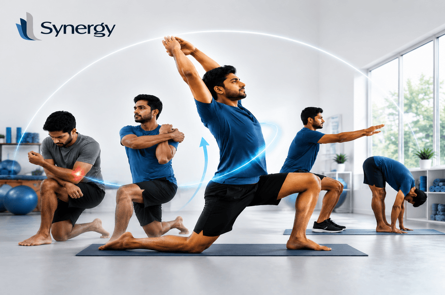
Professional programs for the Flexibility and Mobility Program play a crucial role in your regimen if you desire ease and painlessness in your movements. Regimens that increase the range of motion in joints and increase muscular flexibility are aimed at enhancing posture, physical performance, and functional abilities. This book will introduce you to scientific knowledge concerning the benefits of movement training regimens and explain how to create an ideal body for yourself. The Fears of Being Physically Inflexible Imagine waking up and feeling like you’re walking through thick molasses. When you sit for a long time, and your shoulders hurt, or you feel a pinch in your lower back when you grab your shoes, these aren’t just signs of “getting older.” This normally occurs when the body loses the natural ability to go through its range of motion. At times, flexibility is equivalent to mobility. For example, while you may easily perform hamstring stretches by sitting on the ground, are you able to do so when moving or lunging? At this stage, it is really crucial that you understand the difference between passive stretching and dynamic movement. When our joints get “stuck,” they can make any activity, from going to the gym to playing with your kids, painful and tiring because our muscles have to work harder to make up for it. Learning How the Body Moves To improve our movement, we need to look at the basic parts of physical freedom: mobility and flexibility. Even though they are often used interchangeably, the words mean different physiological processes. What you need to be adaptable If a person’s muscles or groups of muscles can passively lengthen over a range of motion, they are said to be flexible. For example, the “length” of a rubber band. Tendons, ligaments, and fascia are the connective tissues that allow the limb to move. The Effect of Portability The active range of motion of a joint is what makes it mobile. It’s not just how far a joint can go; it’s also how much strength and control you have in that area that matters. A person’s range of motion, strength, and motor control all affect how well they can move around. The goal of a full-body Flexibility and Mobility Program is to make sure your body is both “loose” and strong and stable. How Moving Around Affects Your Health? Moving around is important for all living things. Not only can structured mobility be considered a way to stretch muscles but also a means to reach your nervous system. With the help of movement, we produce synovial fluid which acts as an oil lubricant that keeps our “hinges” from getting worn out and grinding into one another. Consistent mobility drills train the brain to engage with the muscular system, which enhances the effectiveness of the neuromuscular system and reduces the phenomenon called braking, i.e., the inability to move further due to stiff muscles on the opposite side of your movement. Advantages for the cardiovascular system: The secret of holistic health is being healthy both inside and out. Helping your body stay physically fit through consistent mobility exercises, hydration, balanced nutrition, and sufficient sleep plays a huge role. What You Need to Make Your Mobility and Flexibility Program Work? Block of building Main Subject Area Main Benefit Moving while working out Activation of muscles Prepares the heart and muscles for action Reaction to Soap Media (SMR) The release of myofascial Breaks up adhesions in tissues To finish, Isometric Vectors Best performance Makes joints more stable Reduced Joint Movements Health of the joints Keeps the whole joint’s range of motion 1. Flexion Release Techniques To “smooth out” the fascia, or connective tissue that surrounds your muscles, you can use massage balls or foam rollers. This results in fewer “knots” and allows the layers of the tissue to slide over each other freely. 2. Extensive Coordination When doing many activities under pressure, they are considered the most recent techniques for movement training. In modern times, exercises involving dynamic activities that challenge the physical and mental states of an individual have gained popularity as opposed to traditional stretching practices lasting several minutes. Preventing Contemporaneous Diseases The modern way of life is a “flexion” scam. It affects the spine and hips when sitting in a particular posture, looking down at our mobile phones, and driving. When the body adjusts to a specific posture, “normal” motions become challenging. The best cure for this inactive lifestyle is a program focusing on flexibility and mobility. With a strengthened upper back, the individual will reverse the “C-shape” form that leads to headaches and chronic neck pains. Furthermore, by improving the ankle joint range of motion, the knee and lower back complications due to poor foot structure will be solved. Realistic Methods to Start Enhancing Instantly Expert Advice: The Importance of Living Longer In assessing the quality of life when one is elderly, “functional independence” has often been used as a metric among doctors. Your joint flexibility now will tell whether you are capable of stooping down to lift your grandchild or reaching a high shelf. Through investing in these activities now, you are future-proofing yourself against the consequences of ageing. The NHS has stressed the importance of exercising and how it is crucial to have flexibility and balance to avoid accidents and maintain coordination. Similarly, the Mayo Clinic suggests stretching on a consistent basis in order to achieve greater joint flexibility. A Strategy That Will Lead to Overall Wellness: Synergy When many people find themselves trying to balance their hectic life with proper exercise and movement, they often turn to professionals to ensure they move correctly. Since movement is vital in terms of health, the idea of synergy helps to organise this approach. Those who engage in a flexibility program that focuses on enhancing flexibility and mobility will not only begin to move but will also move differently because of the interaction of strength and flexibility. Read this blog from Synergy to learn easy, daily
Movement Therapy for Office Workers to Reduce Daily Pain
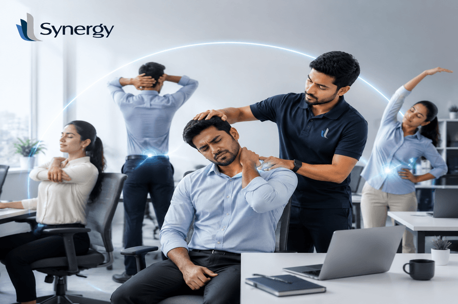
It is quite common nowadays to have chronic pain in our bodies due to the sitting position, which is characterised by diseases such as “tech neck” and lower back problems. Movement therapy for office workers is a must-have tool for anyone who wants to decrease discomfort during working hours and increase workplace productivity. Introduction: Why Your Working Hours Become a Struggle With Tightness At the beginning of your workday at 9:00 AM, you feel quite good, but then around noon time, you start feeling stiffness in your shoulder blades. Then, at 3:00 PM, you begin to automatically massage your neck area, and by the end of the day, when closing your computer, you have an uncomfortable sensation in your lower back as if it were squeezed into one block. This isn’t just “part of the job.” It is a physiological response to static loading—a state where your muscles are held in a fixed position for far too long. When your body remains stationary, blood flow slows down, and the fascia (the connective tissue surrounding your muscles) begins to stiffen. You aren’t just tired; your body is literally crying out for a change in state, which is where movement therapy for office workers becomes essential. Why Movement is the Best Medicine for Your Spine? Static postures, even if you have the most expensive ergonomic chair in the world, lead to what clinicians call “postural fatigue.” Your body was designed for locomotion, not long-term suspension. The inclusion of a movement routine into your life is accompanied by many advantages, clinically speaking: The Core Components of an Office Movement Protocol To truly address the root of the problem, we need to look at the body as a connected chain. Pain in the wrist often starts in the shoulder; pain in the lower back often starts in the hips. 1. Reversing the “C-Curve” of the Spine When you type, your shoulders naturally roll forward and your chin tucks toward your chest. To counter this, you must engage in “extension” exercises. A simple thoracic opener—clasping your hands behind your head and gently leaning back over your chair—can immediately decompress the vertebrae. 2. Addressing “Gluteal Amnesia” Prolonged sitting can lead to your glutes essentially forgetting how to fire, which forces your lower back to take all the strain of your weight. Taking a “standing break” every 30 minutes isn’t just about the legs; it’s about re-engaging the posterior chain. [Link to related blog on postural alignment by Synergy]. 3. Eye and Neck Synchronisation Your neck follows your eyes. If you are squinting at a screen, your head leans forward. Practising the “20-20-20 rule” (looking 20 feet away for 20 seconds every 20 minutes) helps relax the ocular muscles, which in turn reduces tension in the suboccipital muscles at the base of your skull. 4. Dynamic Hip Flexor Releases The psoas muscle shortens when you sit. Over time, this pulls on your pelvis and creates that nagging ache in your lower lumbar region. A simple standing lunge at your desk can lengthen these muscles and take the pressure off your back. How Movement Therapy for Office Workers Creates Lasting Relief? Implementing a structured approach to movement isn’t about gym sessions; it’s about “greasing the groove” throughout the day. Clinical studies suggest that frequent, low-intensity movement is actually more beneficial for metabolic health than one hour of intense exercise followed by eight hours of sitting. By adopting movement therapy, you are essentially training your nervous system to stay alert. This prevents the “slump” that leads to long-term issues like herniated discs or carpal tunnel syndrome. According to research published by the NHS on physical activity, adults should aim to minimise the amount of time spent sitting to reduce the risk of chronic health conditions. Integrating Modern Support with Synergy While movement is the primary tool for recovery, having the right environment to support that movement is equally important. This is where a brand like Synergy becomes a helpful resource for the professional community. It is precisely the expertise of developing a connection between clinical expertise and practical application in the work setting. Synergy doesn’t intend to give a quick fix to people, but rather make some useful suggestions that would inform the users more about their bodies. It is either because of their expertise in ergonomics or perhaps their dedication to integrated well-being that one can look up to them as a mentor for making their health care a professional process. Read about this blog — it tells how small daily habits can prevent long-term pain: Small Changes, Big Relief: Simple Habits for Long-Term Pain Prevention in Daily Life A Healthier Way to Work: Final Thoughts In conclusion, daily pain doesn’t have to be your professional baseline. With knowledge of how static positioning affects muscle fatigue, there is much you can do to regain comfort through movement therapy for office workers. These two benefits alone will make you see the value of this approach. Long-term protection of joints and maintaining a clear mind are the main benefits. Book your posture and movement assessment at Synergy Physiotherapy Clinic today and start working pain-free. Visit Synergy to take the first step toward a healthier workday. FAQS 1. How frequently should I take micro-breaks during my shift? Try to take micro-breaks at least once every 30 to 45 minutes to get back into a normal position and improve blood flow. Even a 60-second posture reset can reduce muscle fatigue significantly. 2. Is movement therapy recommended if I had injuries in the past? Yes, provided you keep movements soft and easy and stop if you have sharp pains. Gentle range-of-motion work can actually support healing when done mindfully. 3. Are there any specific tools needed to execute those stretching exercises? No, as for the most part of the exercises, one needs neither more time nor any other special tools besides a chair. All that is needed is consistency rather than equipment. 4. What can make my neck hurt if I already
Benefits of Strength and Conditioning Rehab for Injury Recovery
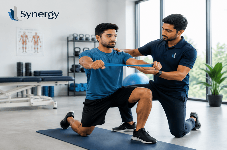
Strength and conditioning rehab forms a bridge between regular physiotherapy treatment and fitness exercises. Although passive treatment may ease the patient’s pain but does not help in regaining the strength of tissues, on the other hand, strength exercise helps the individual to completely recover, regain tissue strength and avoid injuries in the future. Introduction: Beyond the Traditional Approach to Recovery It is natural for the body to react to injury by resting. Traditionally, treatments involved icing, taking pain killers and staying away from any activity. But beyond the traditional approach to healing, it is not pleasant to know that inactivity leads to muscle loss and changes in movement. And here is where strength and conditioning rehab comes in. Different from regular physiotherapy, which may only be about alleviating pain and improving range of movement, this evidence-based practice utilises the very same concepts of progressive overload, periodisation, and multi-joint exercises in a rehabilitative context. In addition to allowing the patient to recover from the existing condition, the practice creates a stronger body better prepared for the future. This particular approach to rehabilitation is applied at Synergy. The clinic’s specialisation in musculoskeletal and orthopaedic rehabilitation allows the therapists to recognise the need for the active involvement of their patients in the process, as well as the concept of proper loading of the recovering parts of the body. Understanding Strength and Conditioning Rehab What Makes It Different? Traditional rehabilitation often follows a linear, cautious path: rest, gentle stretching, basic strengthening, and eventual return to activity. Although effective during acute stages, it may fail to achieve complete functional recovery. The strength and conditioning method, on the other hand, turns the tide. It adopts the same training principles employed by athletes in developing strength and endurance—but in an injured population context. Distinguishing features are presented below: Aspect Conventional Rehabilitation Strength and Conditioning Rehabilitation Loading pattern Minimal load and maximal repetitions Incremental loading according to personal capabilities Type of movement Isolation and unijoint movements Multi-joint and functional movements Progressive advancement Timed progression (Week 2, Week 4, etc.) Criterion progression (Strength goals, pain tolerance, etc.) Purpose Functional restoration Functional restoration plus injury prevention The Science of Progressive Overload The fundamental principle driving this approach is progressive overload—gradually increasing the demands placed on the musculoskeletal system to stimulate adaptation. Research presented at the 2023 American College of Rehabilitation Medicine conference concluded that “progressive overload methods, utilisation of increase in volume, load, and rep scheme adjustments throughout the treatment period, and a strong emphasis on sustainability to avoid re-injury are critical factors in a rehabilitation program”. This is not about pushing through pain. It is about identifying the precise threshold where tissue is challenged enough to adapt without being damaged. Skilled practitioners at Synergy assess your baseline capacity and design a programme that systematically expands your resilience window. Medical Benefits of Strength-Based Recovery 1. Reverses Muscle Atrophy After Immobilisation Within days of joint immobilisation or reduced activity, muscle fibres begin to shrink. This atrophy is not just cosmetic—it directly impairs joint stability and functional capacity. Strength rehab actively counters this decline through targeted resistance exercise. Even more fascinating is the phenomenon of cross-education: training the healthy limb produces strength gains in the immobilised, injured limb through neural pathways . A 2025 review in Sports Medicine – Open confirmed that unilateral resistance training “minimises neuromuscular decline in the injured and immobilised limb” during early rehabilitation phases. 2. Restores Neuromuscular Control Injury disrupts the communication between your brain and muscles. You may have a full range of motion but still feel “unstable” or “off.” This is a neuromuscular deficit—and it is a primary predictor of re-injury. Strength-based rehab retrains these pathways through compound movements that require coordination, balance, and reactive control. Research following ACL reconstruction shows that residual deficits in lower limb performance persist long after standard discharge criteria are met, increasing reinjury risk. 3. Reduces Chronic Pain Through Tissue Capacity Chronic pain often persists not because tissue damage remains, but because the affected area lacks the capacity to handle daily loads. A weak knee hurts when going down stairs. A deconditioned lower back aches after sitting. 4. Prevents Secondary Injuries Compensatory movement patterns develop naturally after injury. You limp. You shift weight. You avoid certain ranges of motion. Such adaptations, though initially protective, soon become habituated and ultimately create other issues in the body, frequently in the opposite extremity or surrounding joints. The re-establishment of symmetry through functional strength rehabilitation allows for breaking the vicious circle. It emphasises bilateral exercises and movement analysis to detect asymmetries prior to becoming new injuries. Synergy’s Approach to Strength and Conditioning Concepts Root Cause Philosophy Synergy is based on an underlying philosophy: the road to recovery is one of faith, competence, and kindness. It means that in the clinic, we go beyond just the obvious symptoms. Every evaluation is comprehensive. Every programme is tailored. And every session is designed to restore not just function but confidence. The Synergy team, led by senior physiotherapists with decades of combined experience, recognises that performance recovery training is not a one-size-fits-all protocol. Your programme might include: Beyond the Injury: Developing Resilience The key difference that defines Synergy is its focus on long-term well-being. As highlighted in their professional profile, the clinic focuses on “long-term healing by addressing issues at the root rather than just managing surface-level symptoms”. This means your discharge plan includes a sustainable strength maintenance strategy—not just a “see you when it hurts again” handshake. At Synergy, located across Anna Nagar, Adyar, T. Nagar, Mogappair, and Vepery in Chennai, that expertise is readily available. For those interested in understanding how movement dysfunction develops in the first place, Synergy has published an insightful blog on Movement Dysfunction Correction Through Targeted Therapy — read this blog, and you’ll know how inefficient movement patterns and compensation can lead to chronic pain and injury, and how targeted correction can retrain your nervous system and improve performance. Conclusion: Reclaiming Your Active Life The days of passive recovery are ending—and
Why Prehabilitation Exercises Matter Before Surgery Recovery
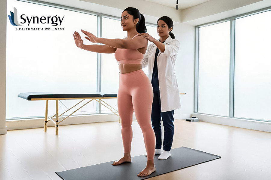
Short Summary Prehabilitation exercises are a planned, physiotherapist-led way to get the body ready for surgery. Patients can improve their chances of a quick recovery by getting ready for it ahead of time by working on their cardiovascular endurance, muscle strength, and joint flexibility. This overview explains why prehab is now required before surgery to make sure it goes well. Intro: Why Prehabilitation Exercises Are Good for Your Body Think of two people who are going to have the same knee replacement surgery. One does nothing but sit around, worry, and not get ready in the weeks before it. By the time one patient gets to the operating room, the other has improved their cardiovascular health, quadriceps strength, and balance through a prehabilitation program supervised by a physiotherapist. Who do you think is responsible for the faster discharge from the hospital? This is a real-life situation. Research from leading orthopaedic institutions shows that patients who don’t take part in pre-surgical conditioning programs are more likely to have problems, take longer to heal, and take longer to become independent again. Still, many people don’t know that there are things they can or should do before surgery. The best time to talk about recovery is right after surgery, when the patient’s body is at its most vulnerable. Prehabilitation turns that way of thinking on its head, and every year more and more data comes in. Prehabilitation exercises is not an afterthought at Synergy Healthcare and Wellness; it is a key part of the surgery process. If you want to change how you deal with your upcoming treatment, you need to know why it matters. Can You Explain What Prehabilitation Exercises Are? Patients go through prehabilitation, or “prehab,” in the days leading up to surgery to get their bodies ready for the physical and mental demands of recovery. It is different from general fitness training because it is focused on the specific condition and is always in line with the type of surgery that is planned. Most of the time, prehabilitation works out three main parts of the body: Prehab includes education in addition to physical therapy. The goal of education is to help patients deal with their worries, know what to expect, and take an active role in their own recovery. This mental preparation, which is often ignored, has an effect on the outcomes of recovery. The Science Behind Why Rehabilitation Works Like other types of physical stress, surgery causes an inflammatory cascade, a short loss of muscle tone (called surgical deconditioning), and a huge rise in energy use to fix damaged tissues. A patient’s capacity to endure surgery is directly correlated with their physical fitness level on the day of the procedure. The British Journal of Anaesthesia says that studies have shown that patients who were more physically fit before surgery were much less likely to have lung problems, stay in the hospital longer, or miss their rehabilitation goals. People who have had joint replacement, heart surgery, or abdominal surgery should pay special attention to this. Some important physiological benefits of structured prehabilitation activities are: Who Benefits from Prehabilitation? Prehab is good for almost all surgery patients, but it has a much bigger effect on the following groups of people: People Who Are Getting Joint Replacements The strength of a person’s core muscles, hip abductors, and quadriceps before surgery has a big effect on how quickly they can walk on their own after knee or hip replacement surgery. If the muscles are stronger before the procedure, they can heal faster. People Who Have Had Ligament Reconstruction Surgery Rehabilitation puts a lot of stress on the muscles around the area, just like when you have surgery to fix a ligament, like an ACL restoration. Improving neuromuscular coordination and strength before surgery makes it easier for the graft to work and for the body to heal. People Who Have Long-Term Illnesses Patients who already have diabetes, obesity, heart disease, or chronic lung disease are more likely to have problems after surgery. Enhancements in glycaemic control, cardiorespiratory fitness, and functional capacity during preoperative rehabilitation can mitigate this risk to some degree. Students in Grades 80 and Up Sarcopaenia, which is the loss of muscle mass that comes with getting older, means that older people will have less strength to work with when they have surgery. A short course of focused prehabilitation exercises can make this balance much less fragile and more resilient during recovery. What Makes a Good Rehabilitation Program Work? Well-designed prehabilitation programs don’t use a one-size-fits-all approach. The plan is tailored to the patient’s current physical condition, the type of surgery being performed, and any other health concerns they may possess. But most evidence-based programs have the following things in common: Part Goal Examples of exercise Exercise power Make the area around the cut stronger. Attacks on the leg, seated knee flexors, and bridges Job that requires physical activity Improve your ability to move before surgery Shoulder rolls, ankle twists, and core exercises Heart health Boosting stamina and heart and lung health Pedalling, riding a bike, and swimming Doing deep breathing exercises Avoid having trouble breathing after surgery. Practice breathing with your lips closed and your diaphragm tight. Body position and sense of balance Make the connection between the muscle and the nerve stronger. Exercises that use a single leg and a wobble board Education and the growth of the mind Set realistic goals and relax your mind. Teaching people how to deal with pain, set goals, and get ready for surgery How Long Does Prehabilitation Usually Last? The best time for prehab depends on the type of surgery and how much time is available before the surgery. Physiotherapy research and clinical guidelines usually say the following: When time is short, any kind of organised preparation is better than none. A physiotherapist might change the intensity and focus of the treatment to make the most of the time that is available. You can find a lot of good advice on rehab
How Gait Training Physiotherapy Improves Walking Recovery
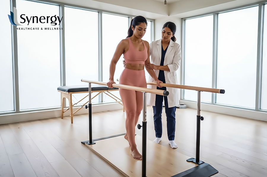
A quick look: Targeted gait training physiotherapy through physical therapy is an important treatment for people who are recovering from neurological disorders, injuries, or surgeries. This particular therapy aids individuals in walking more effectively by realigning their biomechanics, fortifying their muscles, and leveraging neuroplasticity. Structured, evidence-based clinical rehabilitation programs make patients much more independent, help them keep their balance, and lower their risk of falling. The Science of Getting Back to Your Natural Gait: An Introduction to the Reclaiming Movement Many of us don’t realise how complicated the physiological process of walking is until it stops working properly. It can be very limiting when you can’t walk normally, whether it’s because of an injury, a joint replacement, or a neurological event. But you know what? The body is great at learning and changing. Gait training physiotherapy helps people with physical limitations get better by focusing on the complicated mechanics of walking. For a successful recovery, you need to learn how to stabilise your core, evenly distribute your weight, and keep your joints moving at the right speed. This special kind of rehabilitation does more than just “teach” you how to walk again. It changes the way you walk so that it is safer, more effective, and lasts longer. When therapists treat the underlying causes of a patient’s abnormal gait, such as weak muscles or sensory deficits, the patient can walk more confidently and with less pain. 1. The Biomechanics of Gait: What Makes Us Walk in Gait Training Physiotherapy? The stance and swing phases are the two most important parts of a healthy walking cycle. Damage often causes these phases to be uneven. 2. Neuroplasticity and Functional Retraining Neuroplasticity, the brain’s ability to make new connections between neurones, is a fascinating part of physiotherapy for gait training. Training that is both hard and specific to a task can “rewire” the brain to use different pathways to control leg movements when a normal pathway is broken. Clinical Gait Training vs. Regular Exercise Part Full Physical Therapy Rehabilitation for a Bad Walk Listen up Strength and flexibility as a whole Information about how gait works and how stable it is Tools Masseuse and exercise equipment Treadmills, parallel bars, and harness systems Aim Less pain and better overall health Symmetry in stance and freedom of movement The result A body that is more toned A way of walking that is both safe and effective 3. Important Tools and Methods for Rehabilitation Modern gait rehabilitation uses a number of scientifically backed methods to speed up the healing process. (Alt Text :A shot of a female patient using parallel bars for support in a rehabilitation gym, while a male physiotherapist kneels beside her to guide her stepping technique.) 4. Taking a Stand Against Bad Balance and Falling Mental health is a big part of getting better at walking. A rigid stride or “short-stepping” could make it more likely that someone will trip because it makes them less mobile. Clinical studies published on PubMed show that gait therapies that focus on keeping a steady balance can greatly lower the number of falls among older people and people who are recovering from surgery. According to the Chartered Society of Physiotherapy, early movement can help prevent long-term effects like joint stiffness and muscle atrophy. 5. Moving to Mobility in the Real World The goal of every clinical program is to get people out of the gym and onto the street. Part of this process is learning “community ambulation,” which includes things like walking on grass, using curbs, and going up and down stairs. Physiotherapists focus on “dual-tasking” to get their patients ready for the multi-sensory world of everyday life. This means doing two things at once, like walking while holding something or talking. Synergy Physiotherapy: A Place for Clinical Excellence Many clinics offer general help, but real rehabilitation needs a high level of biomechanical accuracy. The main idea behind Synergy is that cutting-edge technology and real-world clinical knowledge work together. Digital gait analysis is an important part of our physiotherapy strategy for gait training. This ensures that your musculoskeletal system is always supported whenever you make any step. We work with people who have difficulties in movement and restore their abilities through comprehensive physical rehabilitation coupled with therapeutic techniques that benefit them. It may be either after undergoing a minor surgery or due to some lifelong problems related to movement. In both cases, we always strive to bring back your “bounce.” This is what we aim for when conducting rehabilitation programs at Synergy – to ensure that people walk in a healthy manner, which is a key characteristic of a youth lifestyle. You can read our previous article on Small Changes, Big Relief: Simple Habits for Long-Term Pain Prevention in Daily Life to understand how everyday routines can have a powerful impact on your physical health. Conclusion: Consistency is the Key to Recovery You need to be careful if you want to get back on your feet. When you do a gait training physiotherapy program, you do more than just move your legs. You also need to reconnect the complicated network that links your brain, muscles, and surroundings. Two of the most important things you can have are patience and determination. You can learn to walk without help if you get the right medical advice and pay attention to how your body moves. A medically-informed plan makes every step toward a more mobile future possible, whether you want to get back to the golf course or just enjoy walking around your garden. FAQ’S 1. How long does it usually take to be able to walk again? Most people say that after four to six weeks of regular treatment, their balance and coordination get better, but results may vary from person to person. 2. Is the physical therapy for gait training painful? When your therapist works with you, they will always think about how comfortable you are. You shouldn’t feel any pain, but you should feel your muscles